
Three Years After MVR: Coco’s Heart Remains Stable
Long-term follow-up of an early mitral valve repair case: mild residual regurgitation, preserved reverse remodeling, and stability at 3 months, 1 year, 2 years, and 3 years
뉴하트동물심장수술센터

Coco underwent regular echocardiographic surveillance for mitral valve disease over several years. Mitral regurgitation (MR) was initially limited, but progressive leaflet degeneration, suspected chordal rupture, and enlargement of the left heart eventually accompanied more pronounced regurgitation. Coco subsequently underwent mitral valve repair (MVR).
Coco belongs to the center’s earlier surgical cohort. Before June 2025, some of our early MVR patients did not have a completely ideal postoperative valve appearance or complete elimination of regurgitation. This case is particularly informative because it demonstrates the long-term course of one such patient rather than focusing on the immediate appearance of the repaired valve alone.
Left atrial size decreased rapidly within three months after surgery. Serial assessments at one, two, and three years documented largely stable regurgitation and cardiac size. At three years, slight additional leaflet degeneration and a small increase in an eccentric MR jet were noted, but left atrial dimensions remained stable and Coco was not receiving cardiac medication.
01. Early Surveillance: Mitral Regurgitation Was Initially Limited
In early 2023, Coco’s MR was not severe. Echocardiography showed an LA/Ao ratio of 1.56, while left atrial and left ventricular dimensions fluctuated around values consistent with ACVIM stage B2. Serial examinations were important to characterize subsequent progression.
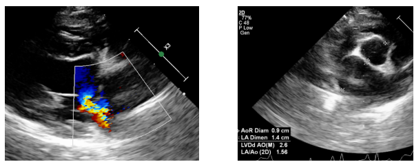
Figure 1. Early color Doppler study showing limited MR. Figure 2. Initial left atrial assessment: LA/Ao 1.56
02. Preoperative Progression: Valve Degeneration and Suspected Chordal Rupture
With time, enlargement of the left atrium and left ventricle became more pronounced. Degenerative changes in the mitral apparatus and echocardiographic findings suspicious for chordal rupture were documented. LA/Ao increased to 2.11, accompanied by substantially more prominent MR on color Doppler imaging. MVR was performed in response to this structural and hemodynamic progression.
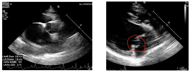
Figure 3. Preoperative left atrial enlargement: LA/Ao 2.11. Figure 4. Abnormal mitral apparatus, with suspected chordal rupture.
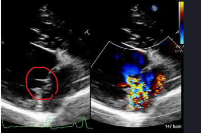
Figure 5. Pronounced preoperative MR and valve changes.
03. Three Months After MVR: Less Regurgitation and Rapid Reverse Remodeling
At the three-month follow-up, only a small amount of residual mitral regurgitation was reported. Left atrial size declined markedly, with LA/Ao reaching 1.27. Compared with the preoperative value of 2.11, this was strong evidence of early postoperative left atrial reverse remodeling.
Right-sided regurgitation remained present and was evaluated separately from the mitral repair. At this point, the TR Doppler velocity was 2.64 m/s with a calculated gradient of 28 mmHg.
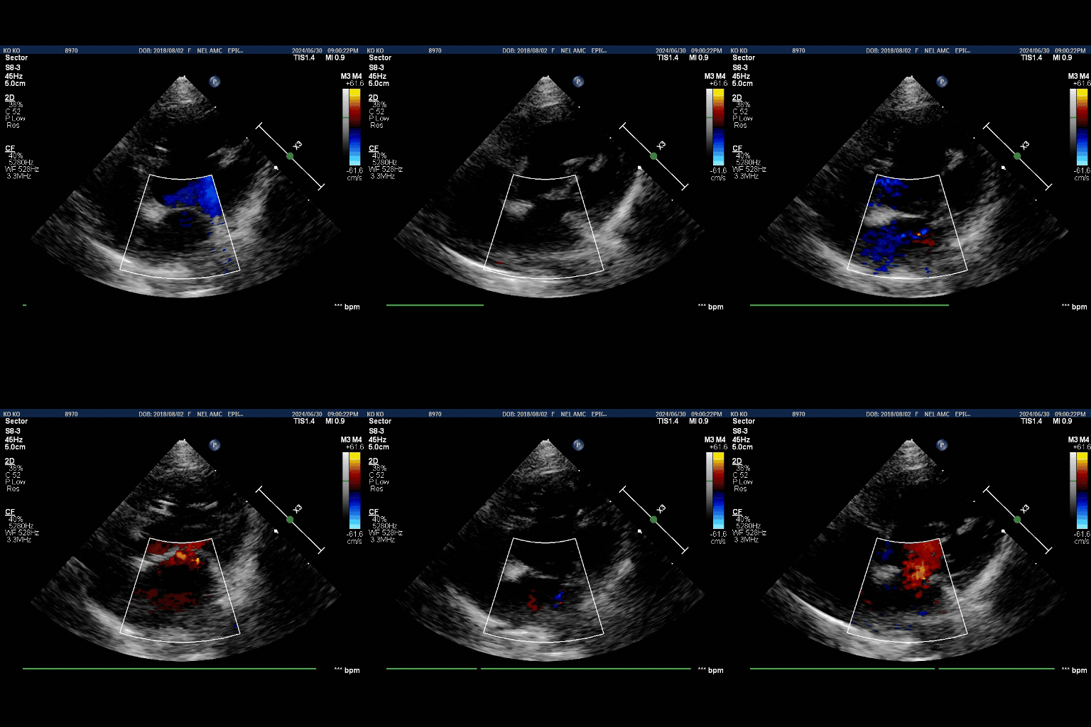
Figure 6. Residual MR at 3 months: representative frame from the original recording.
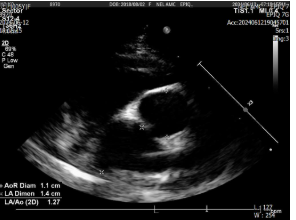
Figure 7. LA/Ao 1.27 at 3 months after MVR.
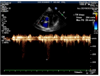
Figure 8. TR Doppler at 3 months after surgery.
04. One Year After MVR: MR, TR, and Left Atrial Size Remained Similar
The appearance of MR on color Doppler was largely unchanged at the one-year follow-up. TR also remained broadly comparable, and LA/Ao was 1.18, showing that the postoperative decrease in left atrial size had been maintained.
One-year TR Vmax was 3.14 m/s (calculated gradient 40 mmHg). A single TR velocity measurement is influenced by hemodynamic conditions and should not, by itself, be treated as proof of progressive right-sided disease.
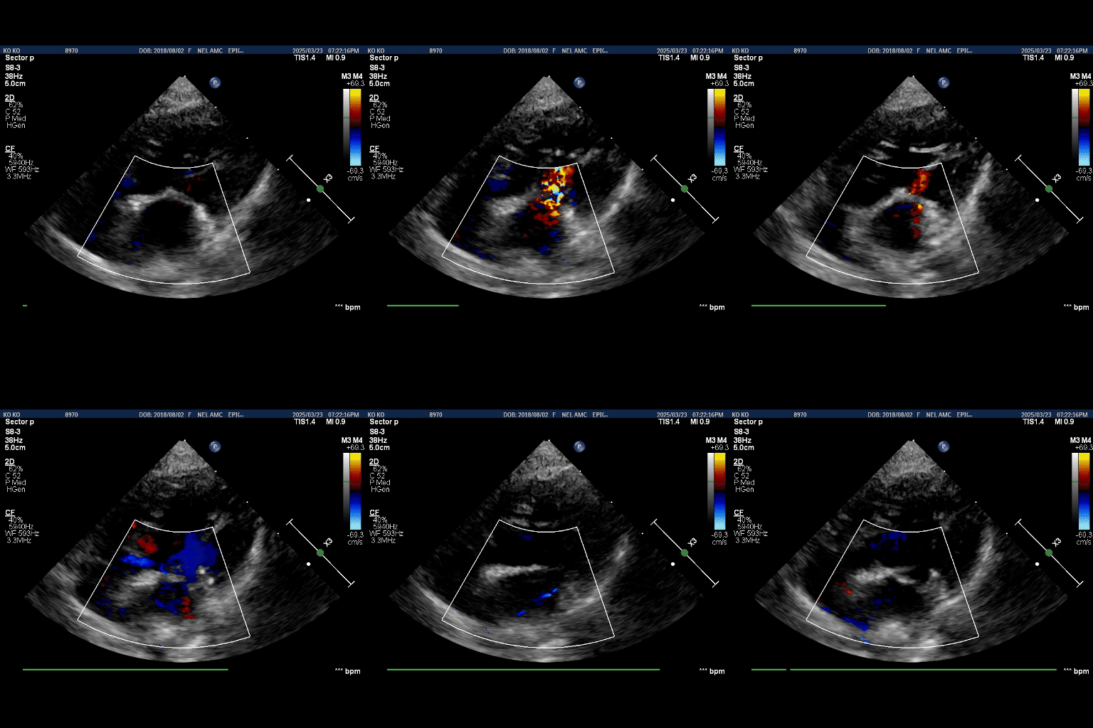
Figure 9. One-year postoperative MR recording.
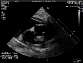
Figure 10. One-year LA/Ao: 1.18.
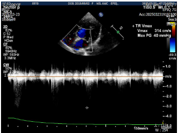
Figure 11. One-year TR: Vmax 3.14 m/s, gradient 40 mmHg.
05. Two Years After MVR: No Clear Recurrent Enlargement
At two years, the color Doppler appearance of MR remained similar to that documented at the prior follow-up. LA/Ao was 1.16, with no evidence in the supplied serial measurements of the marked recurrent left atrial enlargement seen before surgery.
The key question at this stage was not whether the repaired valve had an entirely normal appearance, but whether residual regurgitation was associated with meaningful recurrent cardiac remodeling or clinical deterioration.
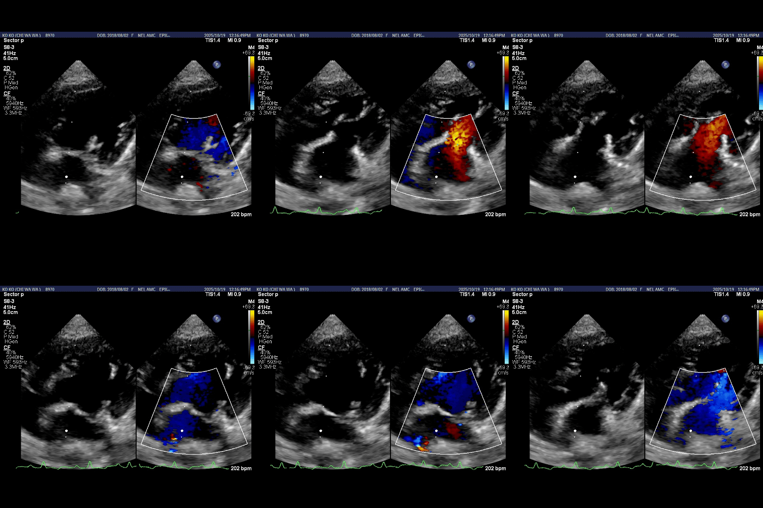
Figure 12. Two-year follow-up: MR remained similar.
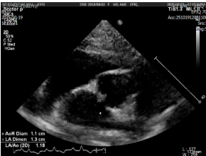
Figure 13. Two-year LA/Ao: 1.16.
06. Three Years After MVR: Minor Valve Changes, but Stable Cardiac Size
At the three-year examination, slight progression of mitral valve degeneration was observed, with a small increase in the eccentric or laterally directed MR jet. However, the change was judged to have limited clinical significance at the time of assessment.
TR also appeared mildly more prominent, but without convincing progression requiring additional cardiac treatment. At three years, TR Vmax was 2.99 m/s with a calculated gradient of 35.77 mmHg; thus the Doppler gradient did not show a sustained increase from the one-year value.
Most importantly, LA/Ao remained 1.24. This was substantially below the preoperative value of 2.11 and did not indicate marked recurrent left atrial enlargement relative to the postoperative measurements at one and two years. Coco currently receives no cardiac medication; ongoing management is directed toward pre-existing hydrocephalus and respiratory problems.
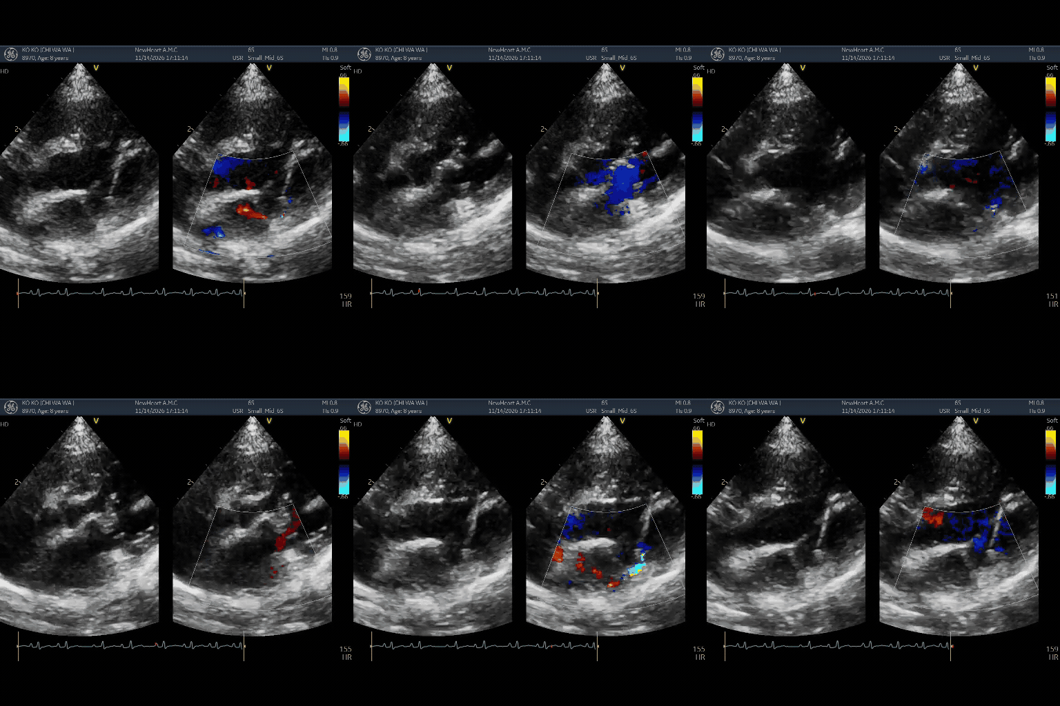
Figure 14. Three-year follow-up: slightly increased eccentric MR.
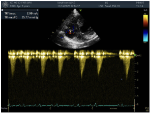
Figure 15. Three-year TR: Vmax 2.99 m/s, gradient 35.77 mmHg.
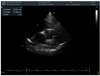
Figure 16. Three-year LA/Ao: 1.24.
07. What Coco’s Case Tells Us About Long-Term MVR Outcomes
This clinical course suggests that the durability of MVR cannot be judged solely by whether the repaired valve is visually perfect or all residual MR has disappeared immediately after surgery. Despite being an early surgical case, Coco demonstrated rapid left atrial reverse remodeling followed by three years without clear recurrent enlargement.
Artificial structures used in mitral valve repair are intended to provide mechanical support for valve coaptation and annular stability. Such structural correction may have contributed to Coco’s durable result. Nevertheless, it does not guarantee that future leaflet degeneration or cardiac enlargement will never occur.
The most clinically relevant observation is the combination of minor MR/TR changes at three years, a stable LA/Ao of 1.24, and continued follow-up without cardiac medication. Serial echocardiographic assessment of MR, TR, leaflet morphology, and left-heart dimensions remains important.
Longitudinal Left Atrial Size: LA/Ao
Preoperative 2.11 → 3 months 1.27 → 1 year 1.18 → 2 years 1.16 → 3 years 1.24
Color Doppler imaging planes and settings may differ between examinations; the colored jet area alone was therefore not used to grade MR quantitatively. The slight progression in leaflet changes and regurgitation at three years reflects the clinical assessment in the supplied record and does not exclude future progression.
References
[1] Keene BW, et al. ACVIM consensus guidelines for the diagnosis and treatment of myxomatous mitral valve disease in dogs. J Vet Intern Med. 2019;33:1127-1140. doi:10.1111/jvim.15488.
[2] Kurogochi K, et al. Long-Term Outcomes of Mitral Valve Repair With Artificial Chordae and Annuloplasty for Myxomatous Mitral Valve Disease in Dogs. J Vet Intern Med. 2025;39:e70171.
NewHeart Animal Cardiac Surgery Center · NEWHART CASE REVIEW
This article is general information and does not replace an individual diagnosis or treatment. If you are worried about your pet, please book a visit.