
PDA Ligation Through an Incision of 3 cm or Less
Kkoji’s surgical correction after two years of age and discharge the following day
뉴하트동물심장수술센터
Kkoji was over two years old and had developed cardiac enlargement associated with a patent ductus arteriosus (PDA). In response to the owner’s preference for a smaller incision, the NewHeart team performed surgical PDA ligation through an incision of 3 cm or less. Postoperative Doppler imaging showed no residual PDA flow, and Kkoji was discharged after a one-day hospital stay.
Hello from NewHeart Animal Cardiac Surgery Center.
In this case review, we share Kkoji’s preoperative thoracic radiographs and echocardiograms, intraoperative photographs of the limited-incision approach, and the clinical and imaging findings observed the day after surgery.
01 A Persistent PDA and Cardiac Enlargement After Two Years of Age
Patent ductus arteriosus (PDA) is a congenital cardiovascular defect in which the fetal vascular connection between the aorta and pulmonary artery remains open after birth. With a typical left-to-right shunt, increased pulmonary blood flow can cause volume overload of the left heart.
Kkoji was already older than two years at presentation. Preoperative thoracic radiographs showed an enlarged cardiac silhouette. Surgical closure was planned to eliminate the abnormal ductal flow and reduce ongoing cardiac loading.
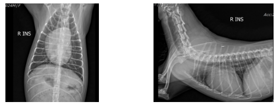
Figure 1. Preoperative ventrodorsal and lateral thoracic radiographs demonstrating an enlarged cardiac silhouette.
02 Distinct PDA Flow on Preoperative Echocardiography
Color Doppler echocardiography clearly demonstrated abnormal flow through the PDA. This examination is important not only for recognizing the anatomic connection, but also for visualizing the location of the shunt and its hemodynamic consequences.
PDA-associated flow was also visualized from the left cranial imaging window. The findings were incorporated into the preoperative assessment and surgical plan.
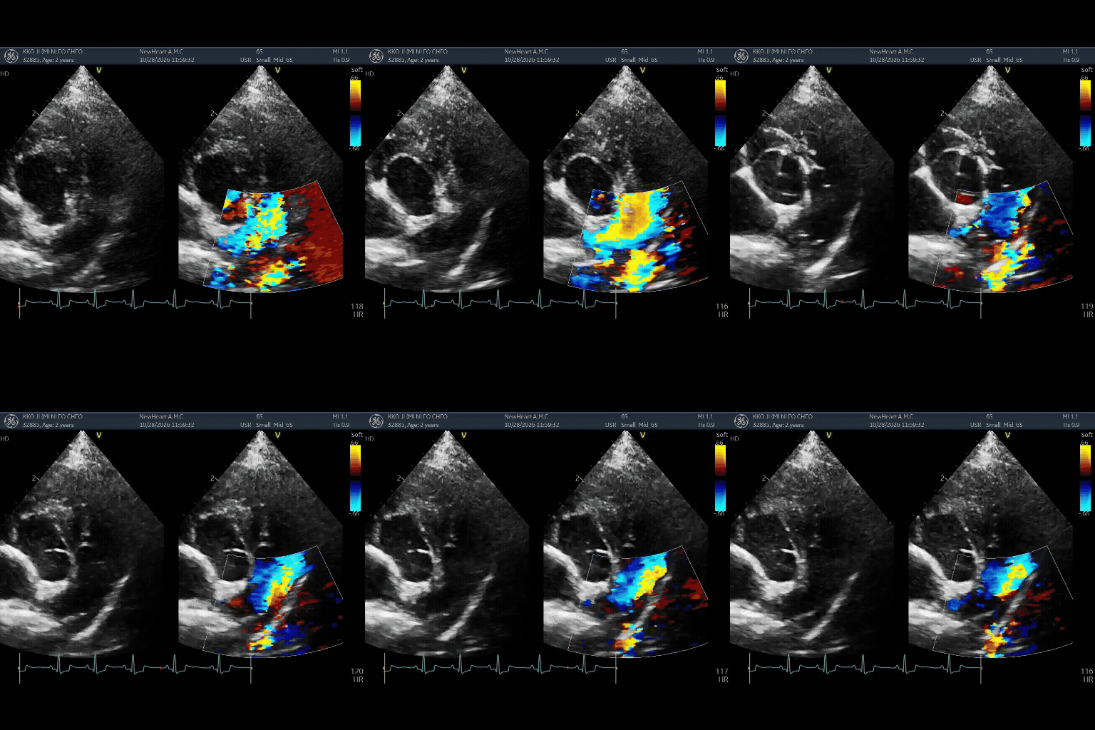
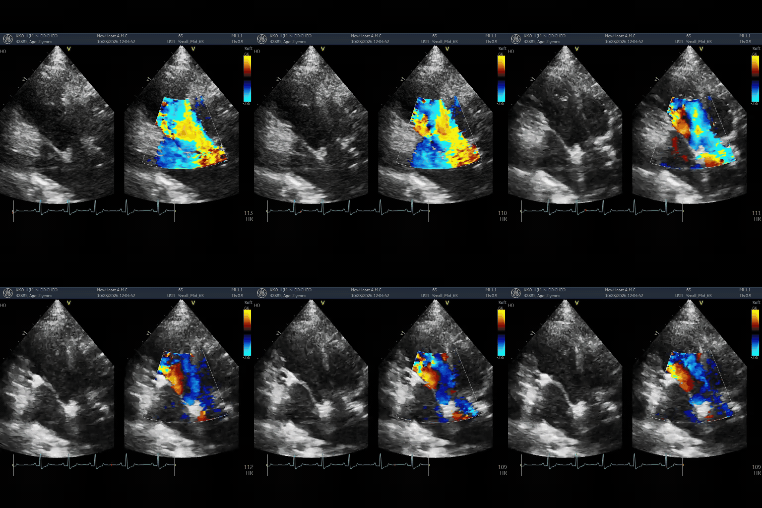
Figure 2. Preoperative echocardiographic images showing PDA-associated color Doppler flow from different imaging windows.
03 Successful PDA Ligation Through an Incision of 3 cm or Less
Kkoji’s owner requested the smallest feasible surgical incision. Considering access to the ductus and intraoperative safety, the NewHeart team used a limited-incision thoracotomy with an incision measuring 3 cm or less.
The ductus was dissected and ligated through the small surgical window, interrupting the abnormal vascular connection. The operative photographs demonstrate the incision size and the confined working field.
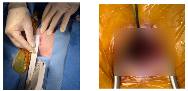
Figure 3. Intraoperative photographs demonstrating PDA ligation through a surgical incision of 3 cm or less.
PDA ligation carries a recognized risk of vascular injury and potentially severe hemorrhage. NewHeart’s experience with open-heart surgery and cardiopulmonary bypass (CPB), together with access to CPB equipment, may provide an additional rescue option when appropriate. However, CPB is not a guaranteed solution to every emergency; feasibility depends on the nature of the complication and the patient’s condition.
04 Postoperative Assessment on the Following Day
Thoracic radiographs were obtained the day after PDA ligation. Together with the clinical findings, the postoperative assessment was consistent with an early reduction in the hemodynamic burden caused by the previously patent ductus.
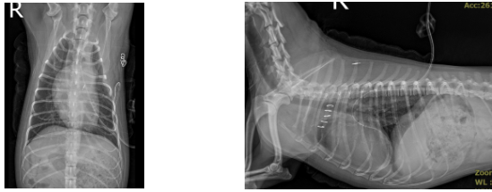
Figure 4. Ventrodorsal and lateral thoracic radiographs obtained the day after surgery to assess postoperative thoracic and cardiac findings.
Postoperative echocardiography was used to reassess cardiac dimensions and function. Elimination of ductal shunting can reduce left-sided volume overload, although the extent of subsequent reverse remodeling requires longer-term echocardiographic follow-up.
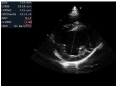
Figure 5. Postoperative echocardiographic image used to reassess cardiac dimensions and loading conditions.
05 No Residual PDA Flow and Discharge After One Day
On postoperative color Doppler examination, the PDA flow observed before surgery was no longer detected. This finding supports successful interruption of the abnormal ductal shunt following ligation.
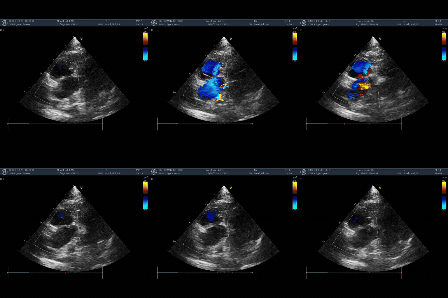
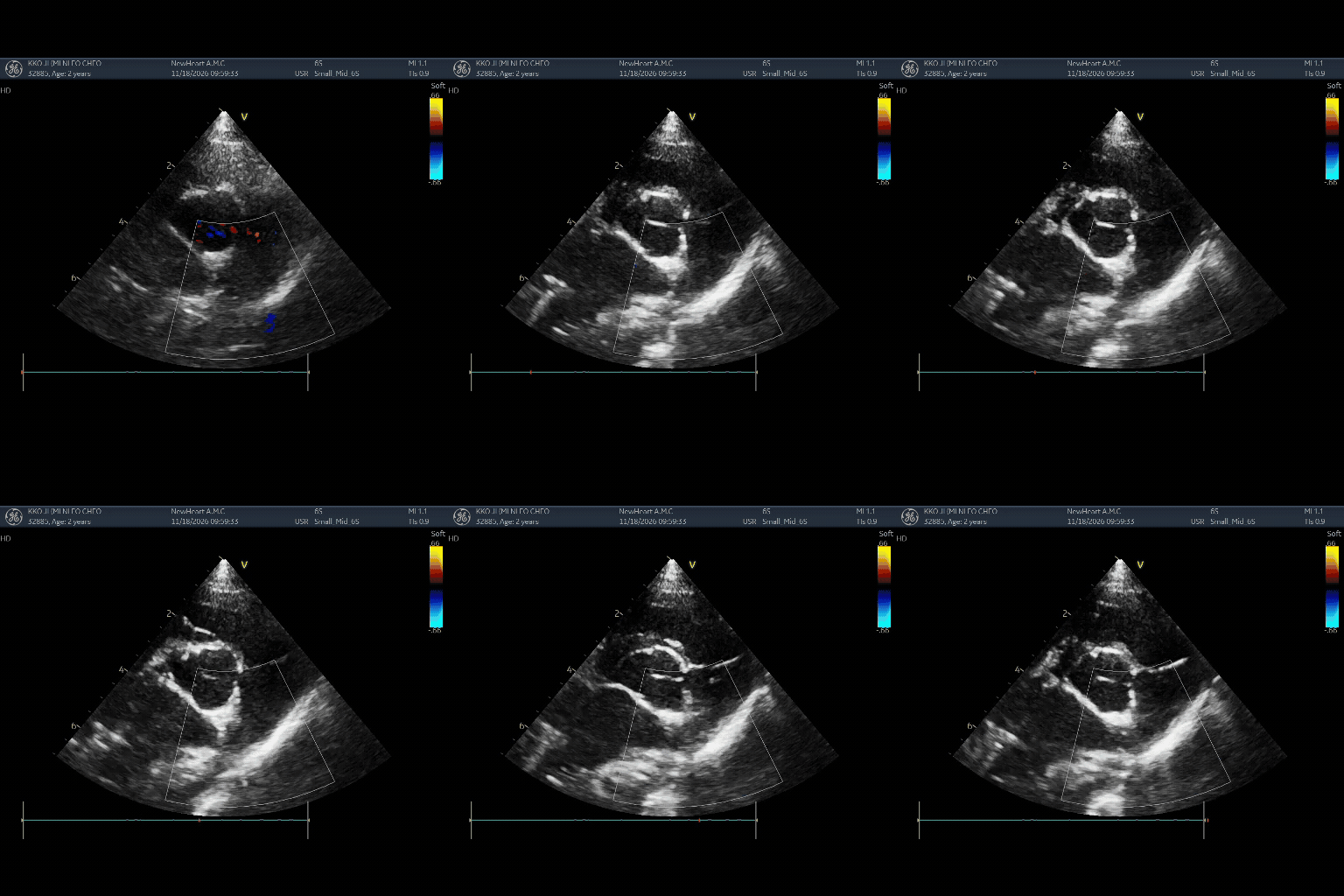
Figure 6. Postoperative color Doppler echocardiography showing no detectable residual PDA shunt flow at the previously identified site.
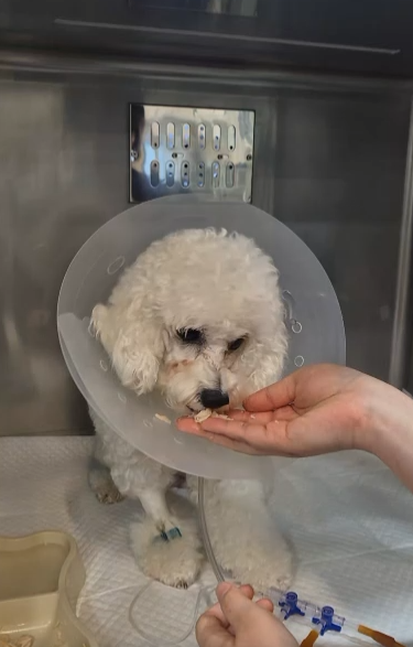
By the following day, Kkoji had a strong appetite and showed no obvious pain response on clinical assessment. With a favorable overall recovery, Kkoji was discharged after one day of hospitalization. This describes this individual case and should not be interpreted as a guaranteed discharge timeline for all patients.
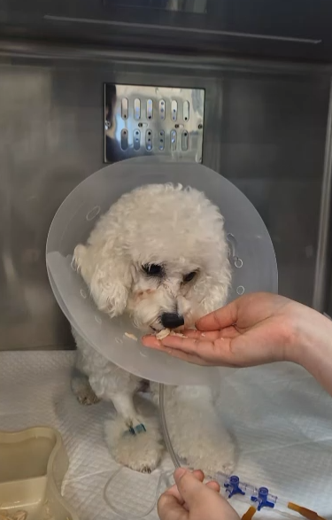
Figure 7. Kkoji during postoperative recovery.
06 Combining a Limited Incision With Surgical Preparedness
Kkoji’s course demonstrates that carefully selected dogs with PDA and established cardiac enlargement may undergo surgical ligation through a limited incision and recover rapidly. Experience in complex cardiac surgery and CPB operations broadens the team’s potential response options in unexpected situations.
Nevertheless, a smaller incision or the availability of CPB does not eliminate surgical risk. Individualized assessment and follow-up for hemorrhage, residual shunting, and cardiac remodeling remain essential.
NEWHART ANIMAL CARDIAC SURGERY CENTER
The technology to repair the heart, the care to continue recovery.
REFERENCES | EVIDENCE
McNamara KL, et al. Risk factors for intraoperative hemorrhage and perioperative complications and short- and long-term outcomes during surgical patent ductus arteriosus ligation in 417 dogs. J Am Vet Med Assoc. 2023. doi:10.2460/javma.22.12.0541.
Grimes JA, Thieman Mankin KM. Surgical ligation of patent ductus arteriosus in dogs: incidence and risk factors for rupture. Vet Surg. 2022;51:592–599. doi:10.1111/vsu.13802.
This article is general information and does not replace an individual diagnosis or treatment. If you are worried about your pet, please book a visit.