

Common heart diseasesWhat each disease is, and how it is treated
From mitral valve disease, the most common heart disease in dogs, to congenital defects and arrhythmia: the conditions NewHeart treats.

- 0%
- 85% of dogs aged 13 or older have this disease.
- yrs
- The most common heart disease in small dogs aged 6 to 7 or older.
It can occur in any breed, but is very common in small, older dogs (Maltese, Poodle, Chihuahua, Shih Tzu, Yorkshire Terrier, Cavalier King Charles Spaniel and others). It usually progresses slowly, and not every dog reaches a severe stage.
What is mitral valve disease?
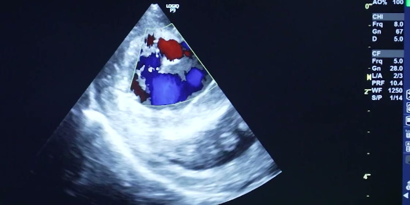
In myxomatous mitral valve disease, the mitral valve between the left atrium and left ventricle thickens and deforms, and blood flows backward.
A healthy valve opens and closes like a door so that blood flows one way. When the valve no longer closes properly, blood leaks back from the left ventricle into the left atrium. Over time the heart enlarges and can progress to heart failure, a state in which the heart cannot do its job.
It is a progressive disease that worsens over time, and it can advance suddenly if a chorda (the cord that holds the valve) ruptures.
What are the signs?
Early MMVD often shows almost no signs, and is frequently first found as a heart murmur. As the disease progresses, the following signs may appear gradually or suddenly.
- Plays less than before and tires easily.
- A dry cough.
- More breathlessness and panting than before. Panting does not settle quickly, and breathing is fast.
- Sits down or faints during a walk or when excited. Stops on walks or asks to be carried.
In severe cases the tongue and gums look blue, and the dog is restless and breathing fast. This is pulmonary edema, fluid in lungs that should hold air, and it is an emergency.
How is it treated?
Unfortunately, no medication slows or stops degenerative change of the mitral valve.

B1
No signs, early stage
- Progress is checked with regular auscultation and echocardiography, once every six months.
- Often it is simply monitored, without medication.
B2
The heart is enlarged, without severe signs
- Early diagnosis by echocardiography and medication can delay progression to congestive heart failure.
- From this stage pimobendan is used to reduce the load on the heart and improve its output.
- Regular check-ups (usually every 3 to 6 months) and lifestyle care (avoiding heavy exercise and obesity) matter.
- In advanced B2, heart surgery can be considered.
C · D
Heart failure with clinical signs
- Congestive heart failure causes serious, life-threatening signs such as breathing difficulty and fainting.
- Several drugs are added to pimobendan. The most important are diuretics such as furosemide or torsemide, with other drugs used to control the disease.
- Combined medication aims to reduce the load on the heart and to prevent or treat pulmonary edema.
- Severe pulmonary edema or breathing difficulty needs emergency care such as oxygen and hospitalization.
- Open-heart surgery under cardiopulmonary bypass directly corrects the damaged valve structure, and in some cases heart medication can be reduced or stopped after surgery.
Other heart diseases
We also treat congenital heart disease and arrhythmia
Pulmonic stenosisPS
- What is it?
- The path from the heart to the lungs is narrowed from birth, putting a heavy load on the right ventricle. The patient tires or becomes breathless with exercise, and may faint in severe cases.
- How is it treated?
Balloon valvuloplasty, an interventional procedure that widens the narrowed area with a balloon catheter, is the standard treatment; surgery may be needed in some cases. Short-headed breeds such as French Bulldogs and Boxers are genetically predisposed.
Atrial septal defectASD
- What is it?
- A congenital hole in the wall between the left and right atria lets blood leak from one atrium to the other. A large hole enlarges the right atrium and ventricle and increases blood flow to the lungs, which can lead to heart failure.
- How is it treated?
Small defects may only be monitored, but large defects or an enlarged heart may need open-heart surgery to close the hole.
Ventricular septal defectVSD
- What is it?
- A congenital hole in the wall between the left and right ventricles. When blood leaks from the left ventricle to the right, blood flow to the lungs increases excessively and the load on the heart grows. It is often found by chance as a murmur.
- How is it treated?
A small VSD may never cause problems, but for a large VSD surgery (patch closure) is considered. Medication is added when heart failure is present.
Aortic stenosisAS / SAS
- What is it?
- The path into the aorta, which carries blood from the heart to the whole body, is narrowed. The left ventricle thickens and the load on the heart grows, bringing a risk of fainting or sudden death.
- How is it treated?
It is mainly managed with medication that reduces the load on the heart; in some severe cases balloon dilation or surgical resection is attempted. Early detection and regular cardiac examinations matter.
Congenital tricuspid valve dysplasiaTVD
- What is it?
- The tricuspid valve between the right atrium and ventricle is abnormal in shape or position from birth, so it closes poorly or is narrowed and the right atrium gradually enlarges. Ascites, coughing and fatigue may appear.
- How is it treated?
Most cases are managed with medication such as diuretics and heart drugs; valve repair surgery is considered in severe cases.
Tetralogy of FallotTOF
- What is it?
- Several congenital defects occur together: typically 1) ventricular septal defect, 2) pulmonic stenosis, 3) an abnormally positioned aorta and 4) right ventricular hypertrophy. The blood lacks oxygen, causing cyanosis (a blue tongue and lips) and exercise intolerance.
- How is it treated?
The anatomy is complex, so several defects must be corrected together by specialist heart surgery, and surgery is not possible in every case. When surgery is difficult or high-risk, medication is used to control the signs.
Patent ductus arteriosusPDA
- What is it?
- A vessel used only before birth (the ductus arteriosus) stays open after birth, so abnormal blood flow continues between the aorta and pulmonary artery. Left untreated, it can become life-threatening through heart failure and pulmonary edema.
- How is it treated?
Complete closure is the standard treatment.
- Surgical ligation of the ductus through a thoracotomy.
- Interventional closure with a coil or device, through a catheter placed in a leg vessel.
Corrected at the right time, the outlook is good.
Arrhythmia that needs a pacemakerPacemaker
- What is it?
- When the heart's electrical signal becomes too slow (severe bradycardia) or is interrupted (for example complete atrioventricular block), the heart cannot beat properly, causing fainting, extreme lethargy and sudden collapse. Some of these cannot be controlled with medication.
- How is it treated?
For life-threatening bradyarrhythmia that will not recover on its own, implanting a pacemaker is the most reliable treatment. A small lead is placed inside the heart and the device under the skin to keep the heart beating at a steady rhythm. Regular device checks and cardiac examinations are needed afterward.