
Removing a Mass That Obstructed the Heart: Ttobo’s Right Ventricular Surgery
Surgical management of an intracardiac mass causing severe right-sided heart failure and right ventricular outflow obstruction
뉴하트동물심장수술센터
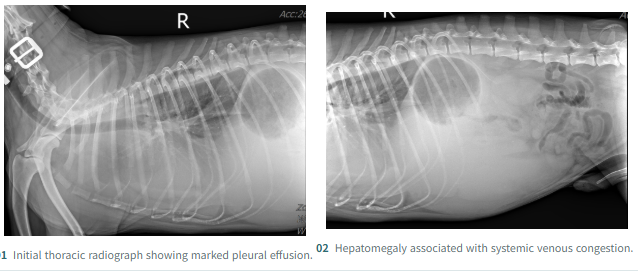
Ttobo presented with difficulty breathing. Diagnostic imaging revealed an intracardiac mass obstructing blood flow from the right ventricle toward the pulmonary artery, accompanied by severe right-sided heart failure. At NewHeart Animal Cardiac Surgery Center, the mass was removed under cardiopulmonary bypass (CPB), followed by serial echocardiographic assessment.
02 A large intracardiac mass compromising right ventricular flow
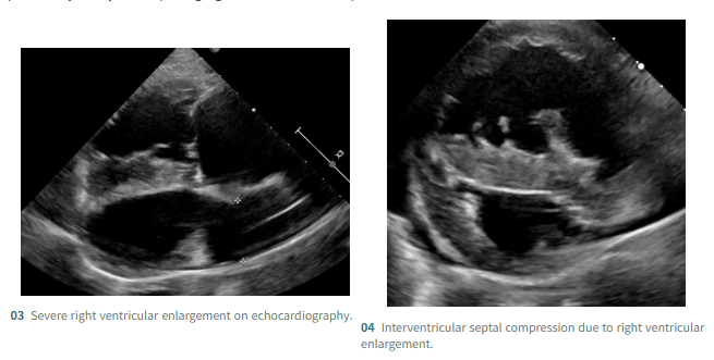
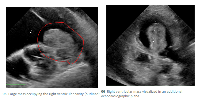
Echocardiography demonstrated substantial right ventricular enlargement with leftward displacement of the interventricular septum. A large mass occupied the right ventricular cavity and interfered with flow toward the pulmonary artery. Tricuspid regurgitation (TR) was also present.
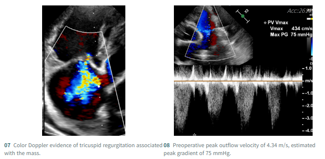
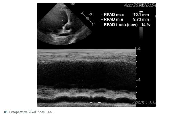
Preoperative Doppler interrogation showed a peak outflow velocity of 4.34 m/s and a corresponding estimated peak gradient of 75 mmHg. The right pulmonary artery distensibility (RPAD) index was 14%, providing an additional indicator for assessment of the right-sided hemodynamic burden.
03 Resection under cardiopulmonary bypass
Resecting a mass inside the right ventricle requires careful management of its relationship to the surrounding cardiac structures and blood-flow pathway. The NewHeart surgical team used CPB, including aortic cannulation, to support intracardiac resection of the mass.
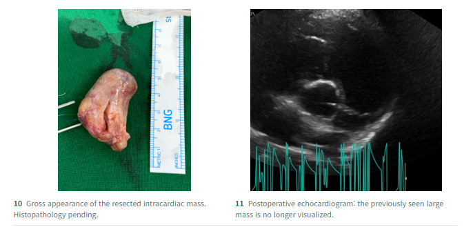
The excised tissue has been submitted for histopathologic examination. The definitive tumor type and its benign or malignant nature remain undetermined. Further assessment of recurrence risk and any additional treatment will depend on the pathology findings.
Histopathology pending | The definitive tumor diagnosis has not yet been established.
04 Postoperative findings: reduced right ventricular distortion
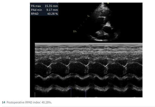
Postoperative echocardiography no longer demonstrated the large mass previously occupying the right ventricular cavity. The right ventricle appeared smaller, and the marked leftward compression of the interventricular septum had clearly decreased. Furthermore, the right ventricular outflow tract obstruction caused by the mass had been successfully relieved, allowing unobstructed blood flow into the pulmonary artery.
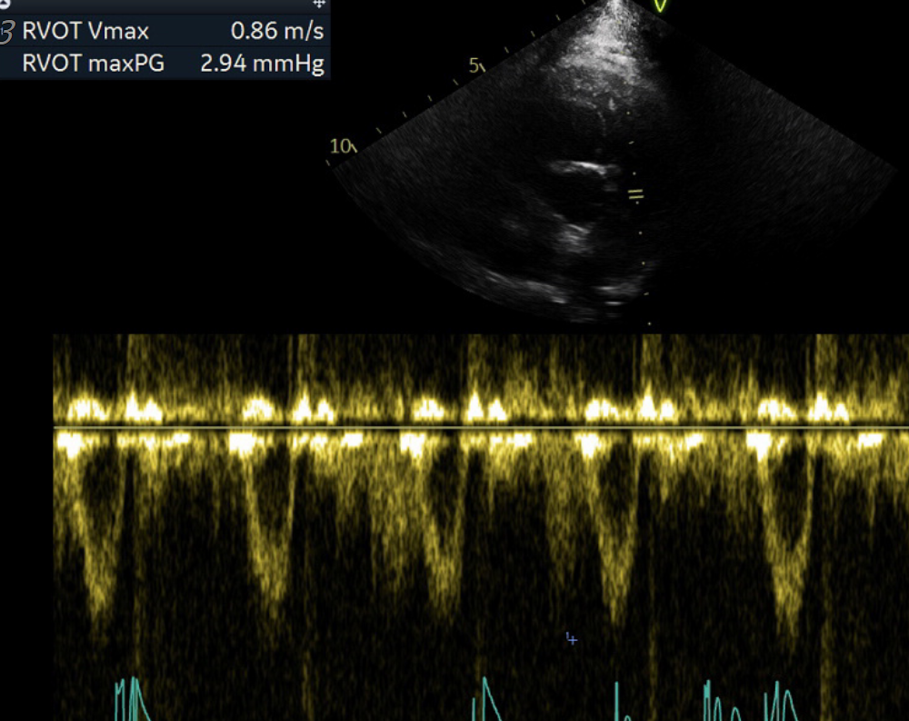
Figure. Resolution of right ventricular outflow tract obstruction (RVOTO).
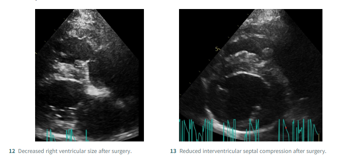
The RPAD index increased from 14% before surgery to 40.28% afterward. This reflects an improvement in the measured pulmonary artery distensibility index, although RPAD alone cannot definitively establish pulmonary arterial pressure or long-term prognosis.
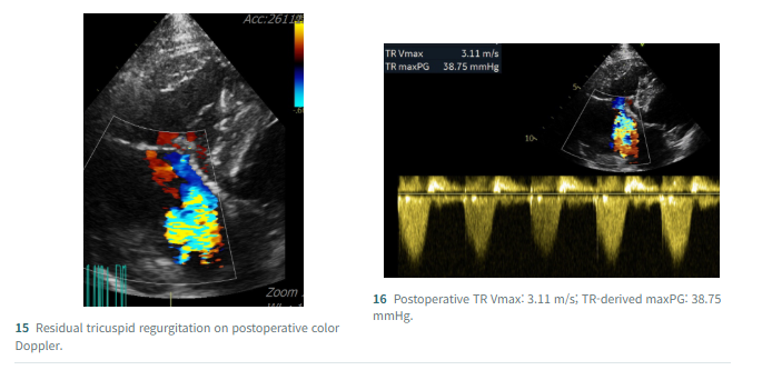
05 Residual tricuspid regurgitation and follow-up care
Tricuspid regurgitation remained detectable after removal of the obstructing mass. Postoperative TR Vmax was 3.11 m/s, with a TR-derived maximum pressure gradient of 38.75 mmHg. These measurements are derived from the tricuspid regurgitant jet and should not be interpreted as the residual right ventricular outflow gradient. Continued assessment of TR, right ventricular dimensions, and signs of venous congestion will be important.
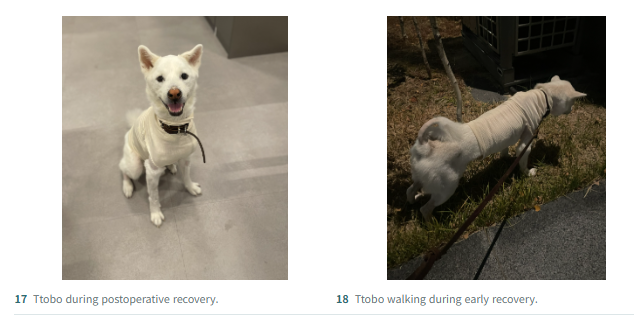
06 Ttobo’s early recovery
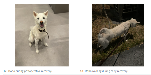
Right ventricular mass resection is technically demanding because surgical access, adjacent cardiac structures, CPB management, and postoperative right-sided hemodynamics must all be considered. NewHeart used an aortic cannulation–based perfusion strategy alongside intensive postoperative care. During early recovery, Ttobo was able to walk and urinate voluntarily.
Removal of the mass was an important step toward relieving the severe obstruction to blood flow. Future care will focus on the histopathology findings, the residual tricuspid regurgitation, and long-term cardiac follow-up. We wish Ttobo a smooth and healthy recovery.
NEWHART | Animal Cardiac Surgery Center
This article is general information and does not replace an individual diagnosis or treatment. If you are worried about your pet, please book a visit.