
Transforming Cardiopulmonary Bypass in Canine Cardiac Surgery: 53 Consecutive Successful Cases of Direct Aortic Cannulation and an International Presentation at the ACVS Surgery Summit
Direct Aortic Cannulation: A Distinctive Surgical Technique at NewHeart
뉴하트동물심장수술센터
Expanding Cardiopulmonary Bypass Strategies for Small-Breed Dogs While Preserving the Carotid Arteries
Canine mitral valve repair (MVR) requires more than technical expertise in reconstructing the mitral valve. Equally important is the ability to safely and effectively manage cardiopulmonary bypass (CPB), which temporarily takes over the functions of the heart and lungs during open-heart surgery.
At NewHeart Animal Cardiac Surgery Center, we have adopted a distinctive approach in which the arterial cannula is inserted directly into the proximal descending aorta rather than the carotid artery.
At the 2026 American College of Veterinary Surgeons (ACVS) Surgery Summit, we presented our experience with 53 consecutive canine MVR procedures performed using this technique, representing the first presentation of this consecutive clinical series. [1]
The key advantages of this approach are ① anatomical preservation of both carotid arteries and ② a perfusion strategy designed to achieve adequate systemic blood flow during cardiopulmonary bypass.

01. Rapid Postoperative Recovery: Serial CRP Results From Our Patients
Postoperative recovery following open-heart surgery should be evaluated not only through visible improvements in activity and overall clinical condition, but also through inflammatory markers and organ function.
Serial C-reactive protein (CRP) measurements from four recent NewHeart patients demonstrated a progressive decline over several postoperative days, eventually reaching the laboratory reference interval of 0–20.
Normalization of CRP is generally considered one of the important indicators supporting discharge readiness at our center, together with clinical stability, cardiovascular function, respiratory status, and other postoperative findings.
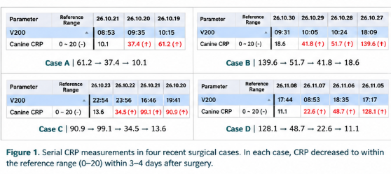
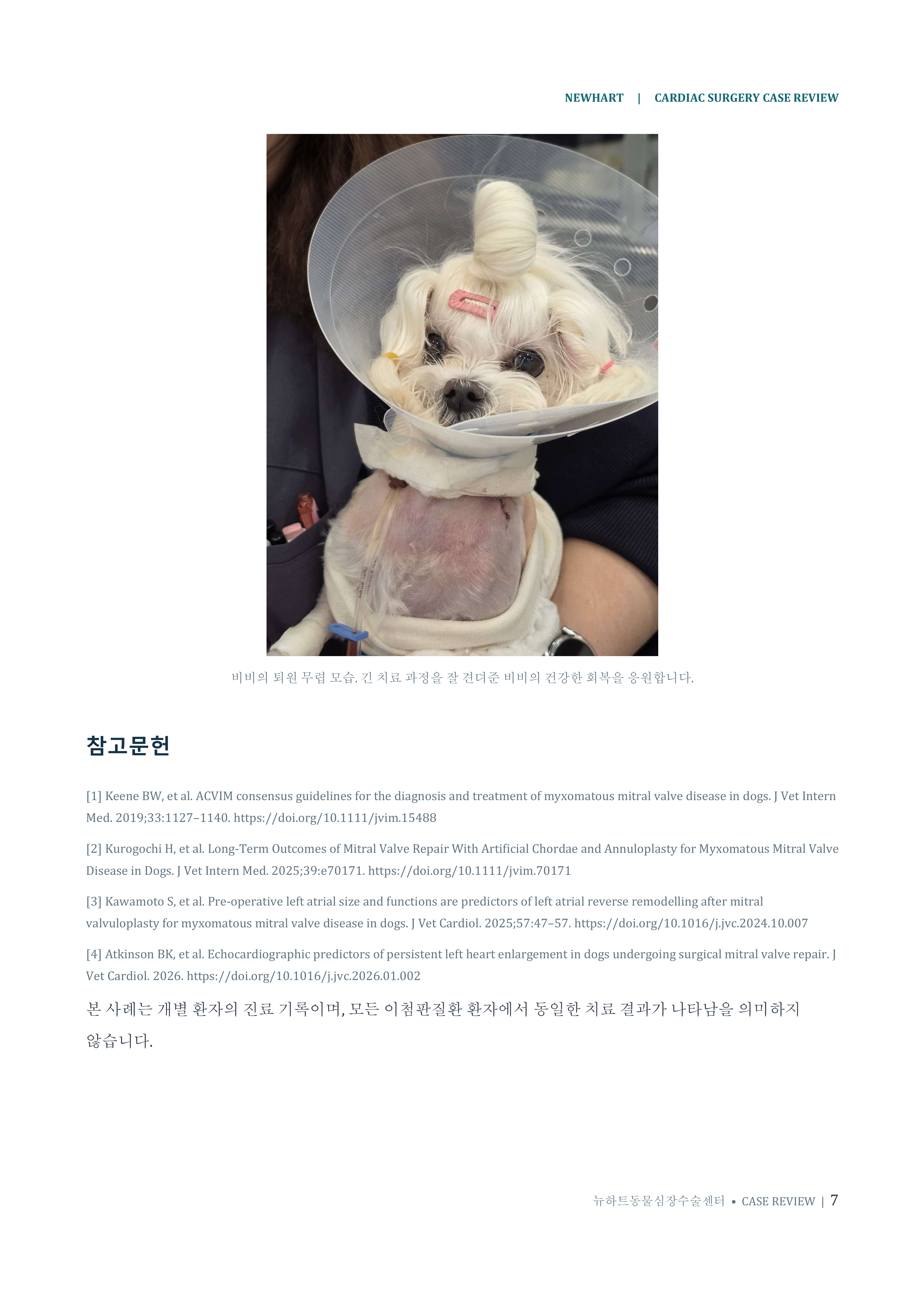
At NewHeart, many patients are able to begin gentle walking within 1–2 days after surgery, and most are discharged approximately 3–4 days postoperatively.
We currently recommend an approximately four-day hospitalization to allow sufficient time for postoperative stabilization, monitoring, and assessment before discharge.
02. Our Experience With 53 Consecutive Cases Presented at the 2026 ACVS Surgery Summit
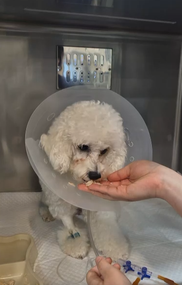
On October 10, 2026, Dr. Taeheum Eom of NewHeart Animal Cardiac Surgery Center presented “Direct Aortic Cannulation for Canine MVR” at the American College of Veterinary Surgeons (ACVS) Surgery Summit. [1]
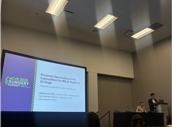
Figure 2. Dr. Taeheum Eom of NewHeart Animal Cardiac Surgery Center presenting the clinical series of direct aortic cannulation at the 2026 ACVS Surgery Summit.
In this consecutive series, direct cannulation of the proximal descending aorta was attempted in 53 dogs undergoing mitral valve repair, and cardiopulmonary bypass was successfully completed using this arterial access route in all 53 patients.
The study population included:
ACVIM Stage B2: 5 dogs
ACVIM Stage C: 32 dogs
ACVIM Stage D: 16 dogs
The median body weight was 3.5 kg (range approximately 1.6–12.5 kg), including extremely small dogs weighing as little as 1.6 kg and patients undergoing repeat cardiac surgery.
Fifty-two of the 53 dogs survived to hospital discharge, corresponding to a discharge survival rate of 98.1%.
This represents an excellent early surgical outcome, comparable to the highest discharge survival rates reported by leading veterinary cardiac surgery centers worldwide.
03. Why the Aorta Instead of the Carotid Artery?
In previously published canine mitral valve repair studies, arterial cannulation through the left carotid artery has been widely described. [2,4]
In small dogs, the limited internal diameter of the carotid artery can impose important technical considerations regarding cannula size, inflow resistance, perfusion pressure, and achievable blood flow.
Direct aortic cannulation reduces the anatomical constraints associated with passing an arterial cannula through a relatively small carotid artery, potentially allowing greater flexibility in establishing adequate systemic perfusion during surgery.
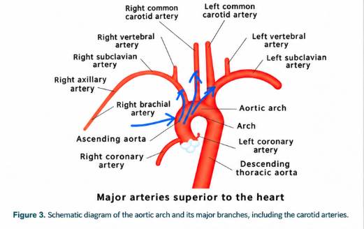
Preserving Both Carotid Arteries
One of the most direct anatomical advantages of aortic cannulation is that carotid artery ligation is not required to establish cardiopulmonary bypass for MVR.
Unlike carotid cannulation techniques that require subsequent vessel ligation, our approach preserves both carotid arteries anatomically and avoids sacrificing a cervical arterial access route that may be useful if repeat surgery becomes necessary.
A study published in 2020, entitled “Striate Artery Infarct After Bilateral Carotid Artery Ligation (BCAL) in a Dog: A Multimodal MRI Study,” documented ischemic cerebral infarction following bilateral carotid artery ligation in a dog. [6]
The study demonstrates that cerebral ischemic injury can occur after carotid artery ligation, despite the presence of collateral cerebral circulation.
Neurological complications are among the important concerns associated with open-heart surgery and cardiopulmonary bypass.
These findings reinforce the potential importance of preserving major arteries supplying the head and brain whenever technically feasible.
However, the reported case involved bilateral carotid artery ligation, and its findings should not be directly extrapolated to the risks of unilateral carotid artery ligation during canine MVR. Further comparative studies are needed to determine whether carotid preservation improves postoperative neurological outcomes.
04. Why Is Direct Aortic Cannulation Technically Challenging?
The technical difficulty of direct aortic cannulation is not simply a matter of inserting a cannula into a larger blood vessel.
The surgeon must simultaneously manage several demanding factors, including the patient's small body size, a restricted operative field, and the continuous movement of the pulsating aorta.
The procedure requires precise aortic dissection, meticulous vascular suturing, and advanced hemostatic techniques.
Successful implementation also depends on seamless coordination among the cardiac surgery team, anesthesiologists, perfusionists, and intensive care specialists.
In dogs weighing approximately 1–3 kg, even a small technical error may result in significant bleeding, vascular injury, or impaired perfusion.
For this reason, direct aortic cannulation requires an experienced multidisciplinary team and carefully standardized surgical and perfusion protocols.
A Technique That Draws International Attention
At international veterinary conferences, one of the questions we frequently receive from veterinary surgeons is:
“Direct aortic cannulation appears technically challenging and potentially risky. How are you able to perform it routinely?”
This question reflects the considerable technical demands of the approach and the expertise necessary to apply it consistently in small-breed dogs.
The ability to perform direct aortic cannulation routinely is one of the distinctive technical capabilities developed by the NewHeart cardiac surgery team.
Because of the demanding vascular techniques, specialized equipment, and extensive team coordination required, this approach may be difficult for other cardiac surgery centers to adopt without substantial training and experience.
05. Perfusion Flow and Temperature: Expanding Cardiopulmonary Bypass Strategies Through Direct Aortic Access
Traditional CPB strategies in small dogs have often combined relatively low perfusion flow rates with deep hypothermia, reflecting the anatomical limitations of small vessels and the technical characteristics of the extracorporeal circulation system.
Direct aortic cannulation may help reduce some of these constraints, providing greater flexibility in maintaining adequate systemic perfusion while performing surgery under moderate hypothermia rather than relying exclusively on deeper hypothermic strategies. [1,2]
A Different Approach to Target Perfusion Flow
In conventional carotid arterial cannulation protocols, target CPB flow rates of approximately 68–80 mL/kg/min have been described.
In contrast, NewHeart generally targets CPB flow rates of at least 100 mL/kg/min when using direct aortic cannulation, with the intention of maintaining adequate systemic perfusion and reducing the potential for hypoperfusion-related organ injury. [5]
These values describe specific perfusion strategies rather than universally applicable physiological limits. The optimal flow rate depends on the patient's condition, body temperature, oxygen delivery requirements, vascular resistance, and CPB circuit characteristics.
Our goal is not merely to achieve a higher numerical flow rate.
Instead, we aim to provide systemic perfusion appropriate to each patient's metabolic and oxygen demands while minimizing unnecessary dependence on profound hypothermia.
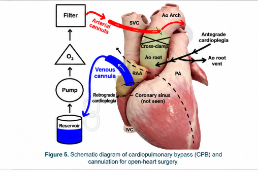
The 2024 EACTS/EACTAIC/EBCP Guidelines on Cardiopulmonary Bypass in Adult Cardiac Surgery, published in 2025, also describe the ascending aorta as a standard arterial cannulation site in human cardiac surgery. [7]
Although the anatomical location and surgical approach differ from NewHeart's proximal descending aortic cannulation technique in dogs, this highlights the established role of direct aortic access in extracorporeal circulation.
06. Why Is Carotid Artery Preservation Particularly Important in Feline Cardiac Surgery?
At NewHeart, preservation of normal vascular structures, including the carotid arteries, is an important consideration not only in canine cardiac surgery but also in complex feline open-heart procedures.
Cats have distinctive cerebrovascular anatomy.
In adult cats, the extracranial segment of the internal carotid artery undergoes substantial developmental regression, while the maxillary artery, arising from the external carotid artery, and its associated rete mirabile play important roles in cerebral blood supply. [8]
Consequently, preservation of the carotid arterial circulation may have particular anatomical and physiological significance in feline patients.
The importance of feline cerebrovascular anatomy has long been recognized.
A relatively recent anatomical review, “Fascinating Wonderful Network: Rete Mirabile of the Maxillary Artery in Cats,” published online in 2023 and in print in 2024, provides a detailed description of the unique arterial network involved in feline cerebral circulation. [8]
These anatomical characteristics support careful consideration of carotid artery preservation when planning feline cardiovascular procedures.
Nevertheless, whether carotid preservation directly improves neurological outcomes or long-term survival in cats undergoing cardiac surgery remains to be established through clinical research.
07. At NewHeart, We Pursue the Best Possible Outcomes — Even When the Path Is Technically Demanding
Following the cases included in our presented clinical series, NewHeart has continued to use direct aortic cannulation as its standard arterial access strategy for open-heart surgery.
Developing and refining this technically demanding procedure required extensive effort, experience, and the ability to overcome numerous surgical challenges.
However, our team has consistently pursued this approach based on the belief that preserving major vascular structures while establishing adequate systemic perfusion represents a valuable direction for advanced veterinary cardiac surgery.
Through repeated clinical application, refinement of surgical techniques, and close collaboration across specialties, we have established a distinctive and highly specialized approach to cardiopulmonary bypass in small animals.
Our experience with 53 consecutive canine MVR procedures, a 100% technical success rate for the planned arterial cannulation approach, and a 98.1% discharge survival rate demonstrates the feasibility of this strategy in a challenging population of small-breed dogs.
NewHeart Animal Cardiac Surgery Center remains committed to evaluating systemic perfusion, postoperative organ function, recovery, and long-term clinical outcomes through continued research and clinical follow-up.
We believe that systematically analyzing these outcomes will contribute to improving the safety and effectiveness of open-heart surgery in both dogs and cats.
Our mission is not simply to develop advanced surgical techniques, but to continuously refine every aspect of perioperative care in pursuit of the best possible outcomes for our patients.
NEWHART ANIMAL CARDIAC SURGERY CENTER
The Expertise to Repair the Heart.
The Perfusion Technology to Protect It.
This article is general information and does not replace an individual diagnosis or treatment. If you are worried about your pet, please book a visit.