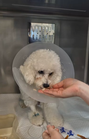

Feline Hypertrophic Obstructive Cardiomyopathy (HOCM): Moji’s Surgical Case
Eight-month follow-up after partial myectomy in a cat with recurrent pulmonary edema and severe left ventricular outflow tract obstruction.
뉴하트동물심장수술센터
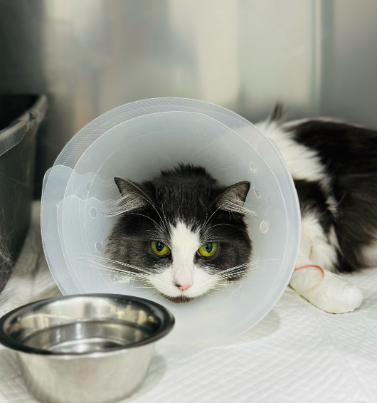
A pioneering feline HOCM surgery: How did Moji’s heart change over eight months?
Eight-month follow-up after partial myectomy in a cat with recurrent pulmonary edema and severe left ventricular outflow tract obstruction.
After the first episode of pulmonary edema in November 2023, Moji received long-term medical treatment for heart failure. Despite treatment, episodes of pulmonary edema accompanied by respiratory distress recurred. Cardiac imaging also revealed severe narrowing of the pathway through which blood leaves the left ventricle and enters the aorta. Following extensive discussion with the owner, the NewHeart cardiac surgery team elected to address the obstruction surgically, as medical management alone had not adequately controlled the underlying hemodynamic problem.
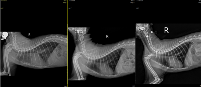
Figure: Progressive cardiac enlargement on serial thoracic radiographs from 2023 to 2025.
This long-term case review focuses not only on the immediate surgical outcome but also on changes in echocardiography, thoracic radiography, and cardiac biomarkers during the subsequent eight months.
01. What is the difference between HCM and HOCM?
Feline hypertrophic cardiomyopathy (HCM) is characterized by thickening of the left ventricular myocardium and may impair ventricular relaxation and filling. Hypertrophic obstructive cardiomyopathy (HOCM) refers to a form in which the left ventricular outflow tract (LVOT) becomes narrowed during systole. Septal and papillary muscle hypertrophy, their position and morphology, and systolic anterior motion (SAM) of the anterior mitral leaflet may contribute to the obstruction. [1]
Severe outflow obstruction can increase the pressure the left ventricle must generate to eject blood and may be associated with SAM-related mitral regurgitation. However, not every cat with HCM develops obstruction, and the presence of obstruction alone does not determine whether heart failure will occur or predict long-term prognosis. [1]
02. Recurrent pulmonary edema and the limitations of medical treatment
Since Moji’s first pulmonary edema episode in November 2023, medical treatment had been adjusted repeatedly. Nevertheless, pulmonary edema recurred, and preoperative examinations revealed marked papillary muscle hypertrophy and substantial LVOT flow disturbance. Renal dysfunction subsequently developed during prolonged heart failure treatment, raising concern for a cardiorenal interaction.
Diuretic-based medical management remains central to the treatment of feline congestive heart failure. In Moji’s case, however, the increasing need for diuretics alongside worsening renal function raised serious concerns about the sustainability of continued medical treatment. [1]
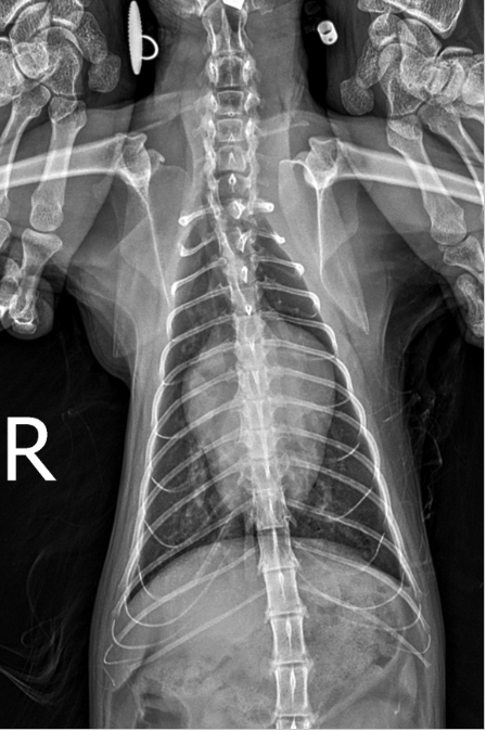
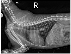
Figure 1. Preoperative thoracic radiograph.
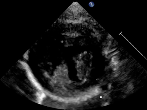
Video 1. Preoperative papillary muscle hypertrophy.
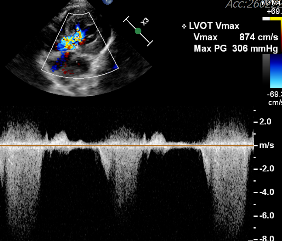
Severe aortic outflow tract obstruction before surgery.
03. Creating more space in the obstructed outflow tract: partial myectomy
In humans with symptomatic obstructive HCM, septal myectomy can be performed at experienced centers when severe symptoms persist despite appropriate medical treatment. The purpose is not simply to make the myocardium thinner, but to enlarge the LVOT and relieve hemodynamic stress related to obstruction and SAM. The 2024 US guideline for HCM recognizes surgical septal reduction therapy as an important option in appropriately selected human patients. [2]
The feline heart differs substantially from the human heart in size and anatomy, and large clinical studies establishing the safety and efficacy of this approach in cats are unavailable. For Moji, the decision to proceed reflected the individual combination of severe obstruction and recurrent heart failure, following a detailed discussion of these uncertainties with the owner.
During cardiopulmonary bypass, a portion of the hypertrophied myocardium was resected using a partial myectomy technique. The aim was to widen the passage from the left ventricle to the aorta and reduce hemodynamic burden. The early postoperative recovery was challenging, but Moji recovered and proceeded to scheduled long-term follow-up.
04. What changed during the eight months after surgery?
1. LVOT: no evident recurrence of severe obstruction
The outflow disturbance evident on the preoperative study was markedly reduced at the eight-month follow-up, without clear evidence of recurrent severe LVOT obstruction.
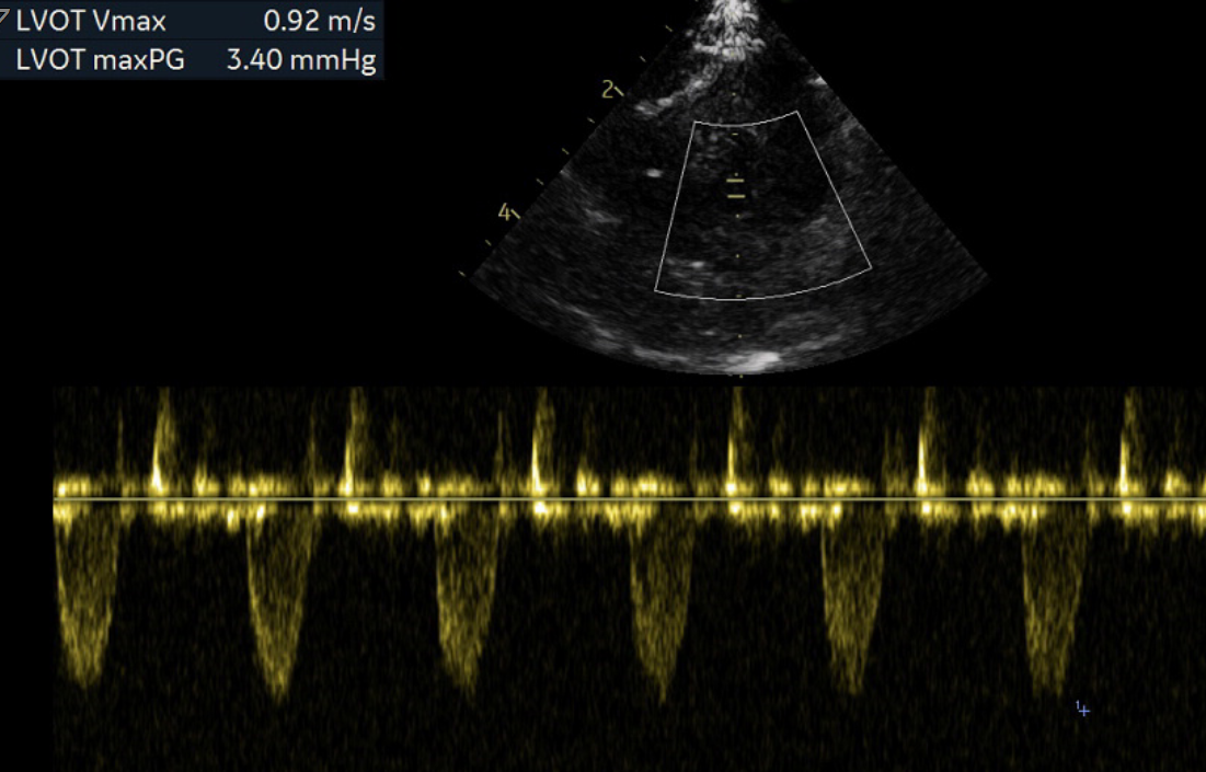
Figure 2. LVOT obstruction before surgery and its appearance after surgery.
2. Myocardium, papillary muscles, and left atrium: changes consistent with reverse remodeling
Myocardial and papillary muscle thickening observed before surgery appeared reduced at eight months. Follow-up echocardiography also showed a decrease in left atrial size. These findings are consistent with reverse remodeling following a reduction in cardiac loading conditions. However, a single case cannot establish the relative contribution of surgery, the natural course of disease, and subsequent treatment changes.
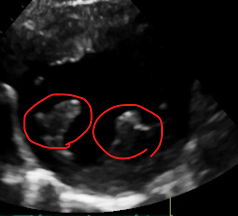
Figure 3. Papillary muscle morphology before and after surgery.
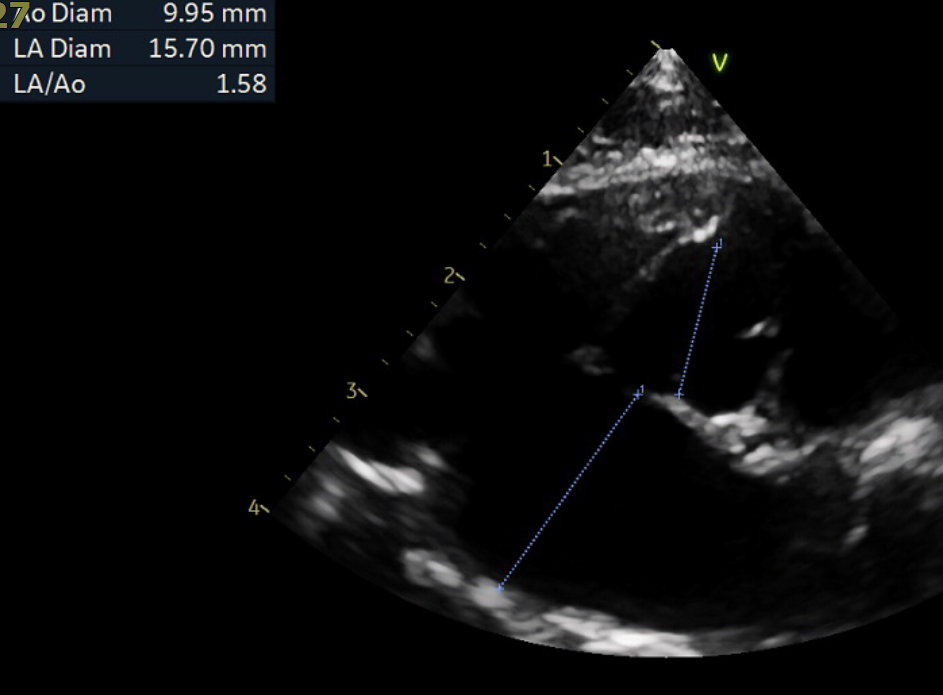
Figure 4. Left atrial size after surgery at eight months.
3. Thoracic radiographs: progressively smaller cardiac silhouette
Serial thoracic radiographs obtained in April, May, and October showed a progressive reduction in cardiac silhouette size. Together with the echocardiographic findings, these images suggested a consistent pattern of improvement.
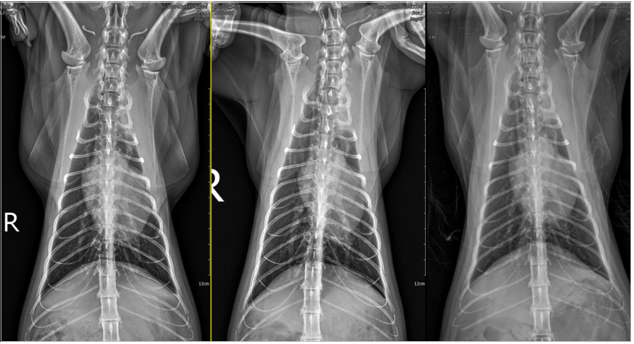
Figure 5. Serial thoracic radiographs obtained in 2,3,8 month after surgery
4. NT-proBNP: improvement during follow-up
The serial NT-proBNP results showed an overall decline after surgery, following a period of progressive elevation before the intervention. As with any cardiac biomarker, the trend should be interpreted alongside the clinical findings and echocardiographic results. A decline in NT-proBNP does not, by itself, establish complete resolution of myocardial disease.

Serial NT-proBNP measurements before and after surgery.
5. No cardiac medications at the eight-month follow-up
At eight months after surgery, Moji was living without cardiac medications. For a cat that had experienced recurrent pulmonary edema and required prolonged medical treatment, this represents a particularly meaningful clinical change. However, discontinuation of medication should not be equated with a cure of the underlying cardiomyopathy. Ongoing monitoring for recurrent obstruction, myocardial hypertrophy, and left atrial changes remains necessary.
05. What does this case mean, and what questions remain?
Moji’s case demonstrates the potential applicability of a surgical principle established for human obstructive HCM to a carefully selected feline patient, with favorable functional and morphological findings persisting through eight months of follow-up. It provides a direction for further investigation rather than proof that the procedure is suitable for all cats with HOCM.
Why NewHeart is documenting Moji’s case
Moji’s outcome should not be interpreted as a recommendation that every cat with obstructive hypertrophic cardiomyopathy undergo surgery. Rather, it illustrates what may be possible for one cat with recurrent heart failure and severe obstruction despite conventional management, when careful patient selection is combined with cardiac surgery, anesthesia, cardiopulmonary bypass, and intensive postoperative care.
The NewHeart Animal Cardiac Surgery Center plans to continue monitoring Moji’s respiratory status, daily activity, LVOT flow, left atrial dimensions, myocardial thickness, and cardiac biomarkers. We hope that the favorable findings persist and that the accumulated clinical results can be assessed and shared academically.
This article is a case review based on an individual patient’s medical records. Appropriate treatment varies according to disease morphology, progression, and comorbidities.
References
[1] Luis Fuentes V, et al. ACVIM consensus statement guidelines for the classification, diagnosis, and management of cardiomyopathies in cats. J Vet Intern Med. 2020;34:1062–1077. doi:10.1111/jvim.15745.
[2] Ommen SR, et al. 2024 AHA/ACC/AMSSM/HRS/PACES/SCMR Guideline for the Management of Hypertrophic Cardiomyopathy. Circulation. 2024;149:e1239–e1311. doi:10.1161/CIR.0000000000001250. (Evidence from human medicine.)
[3] Buchanan K, et al. Resolution of dynamic left ventricular outflow tract obstruction and reversible left ventricular hypertrophy in 4 cats. Can Vet J. 2024;65:1028–1033. PMID:39355694.
[4] US FDA. FDA Conditionally Approves Drug for Management of Ventricular Hypertrophy in Cats. March 14, 2025; Felycin-CA1 official prescribing information. (Indicated for subclinical HCM; excludes cats with current or previous CHF.)
[5] Rivas VN, et al. Cardiac myosin inhibitor, CK-586, minimally reduces systolic function and ameliorates obstruction in feline hypertrophic cardiomyopathy. Sci Rep. 2024. PMID:38802475. (Experimental pharmacologic evidence.)
[6] Cleveland Clinic, Consult QD. When Hypertrophic Obstructive Cardiomyopathy Presents with Barlow’s Valve: A Case Study in Myectomy. 2019. (Human surgical case and case-review structure.)
This article is general information and does not replace an individual diagnosis or treatment. If you are worried about your pet, please book a visit.