
From Respiratory Arrest to Recovery: Gyeoul’s Mitral Valve Repair
A canine case of rapid progression to pulmonary edema, resuscitation, and discharge after open-heart surgery
뉴하트동물심장수술센터
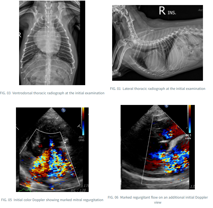
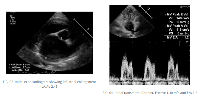
At the initial consultation, Gyeoul was not considered to be in an immediate cardiac emergency. Nevertheless, echocardiography had already revealed marked left atrial enlargement (LA/Ao 2.45) and substantial mitral and tricuspid regurgitation.
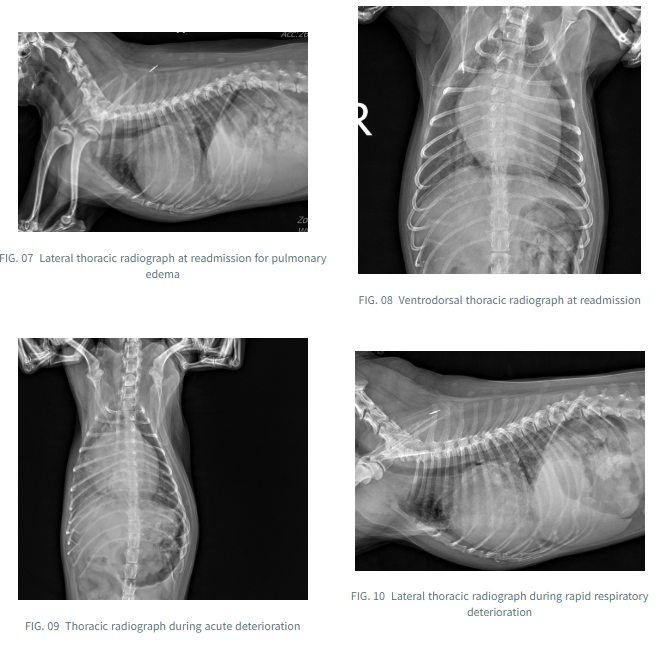
Approximately one month later, Gyeoul returned with cardiogenic pulmonary edema. Respiratory status deteriorated dramatically over the next several hours, culminating in respiratory arrest requiring cardiopulmonary resuscitation (CPR). Repeat echocardiography suggested additional chordal rupture with severe mitral regurgitation. Following discussion with the owner, mitral valve repair (MVR) was performed.
This case review follows the rapid preoperative deterioration and the echocardiographic and radiographic changes documented at hospital discharge.
01 Initial Assessment: Substantial Remodeling Before Clinical Crisis
At the first examination, Gyeoul was not in an immediate state of decompensated heart failure, although the heart was enlarged and coughing was prominent. Thoracic radiographs and echocardiography demonstrated marked enlargement of the left atrium.
An LA/Ao ratio of 2.45 indicated substantial left atrial enlargement. Both mitral and tricuspid regurgitation were prominent, and the transmitral E-wave velocity was 1.40 m/s. This Doppler measurement should be interpreted alongside the remaining imaging findings and clinical context; it does not independently establish left atrial pressure.
02 One Month Later: Pulmonary Edema and Respiratory Arrest
Approximately one month after the initial visit, Gyeoul was readmitted for pulmonary edema. Although the initial presentation was not maximally critical, respiratory function deteriorated dramatically within hours, progressing to respiratory arrest and the need for cardiopulmonary resuscitation.
This rapid course illustrates how abruptly clinical stability can change in a patient with significant valvular disease. More than one factor may contribute to such deterioration, and no single imaging feature can fully explain the entire course.
03 Reassessment: Suspected Additional Chordal Rupture and Severe MR
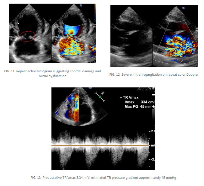
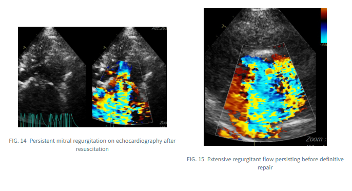
Repeat echocardiography identified substantially worse mitral regurgitation, with abnormal leaflet motion suggestive of additional chordal rupture. Extensive regurgitation persisted even during management after resuscitation.
Before surgery, TR Vmax was 3.34 m/s, corresponding to a pressure gradient of approximately 45 mmHg. This finding contributes to assessment of the probability of pulmonary hypertension, but does not establish its presence or severity in isolation.
Given the suspected structural deterioration of the valve, persistent severe regurgitation, and rapidly worsening clinical condition, surgical intervention was discussed with the owner, and mitral valve repair (MVR) was undertaken.
04 At Discharge: Improved Valve Competence and Left Atrial Size
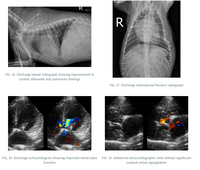
The discharge echocardiogram showed improved mitral leaflet coaptation and function, with no significant residual mitral regurgitation on color Doppler. Thoracic radiographs also demonstrated improvement in the postoperative cardiac and pulmonary findings.
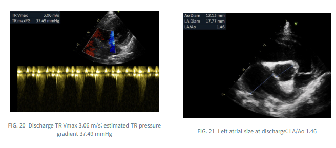
Left atrial size decreased from LA/Ao 2.45 to 1.46, consistent with relatively rapid reverse remodeling after correction of the valvular lesion.
At discharge, TR Vmax was 3.06 m/s, with a corresponding gradient of 37.49 mmHg, lower than the preoperative measurements. However, a lower TR velocity alone does not prove complete resolution of pulmonary hypertension, and follow-up assessment remains important.
05 What Gyeoul’s Case Illustrates
Gyeoul did not appear to be in an immediate life-threatening crisis at the initial visit, but experienced pulmonary edema, respiratory arrest, and resuscitation approximately one month later. Following surgical repair of the structural mitral valve abnormality, the discharge examination documented minimal residual regurgitation and substantially reduced left atrial size.
This individual case does not imply that all dogs with mitral valve disease require surgery. Instead, it highlights the value of repeated echocardiographic assessment and carefully individualized surgical decision-making when a patient deteriorates rapidly.
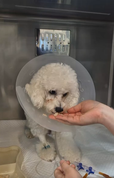
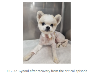
We wish Gyeoul continued recovery and a healthy life ahead following this extraordinary challenge.
This case review describes the documented course through hospital discharge. It does not establish long-term outcomes or imply that identical results can be expected in other patients.
This article is general information and does not replace an individual diagnosis or treatment. If you are worried about your pet, please book a visit.