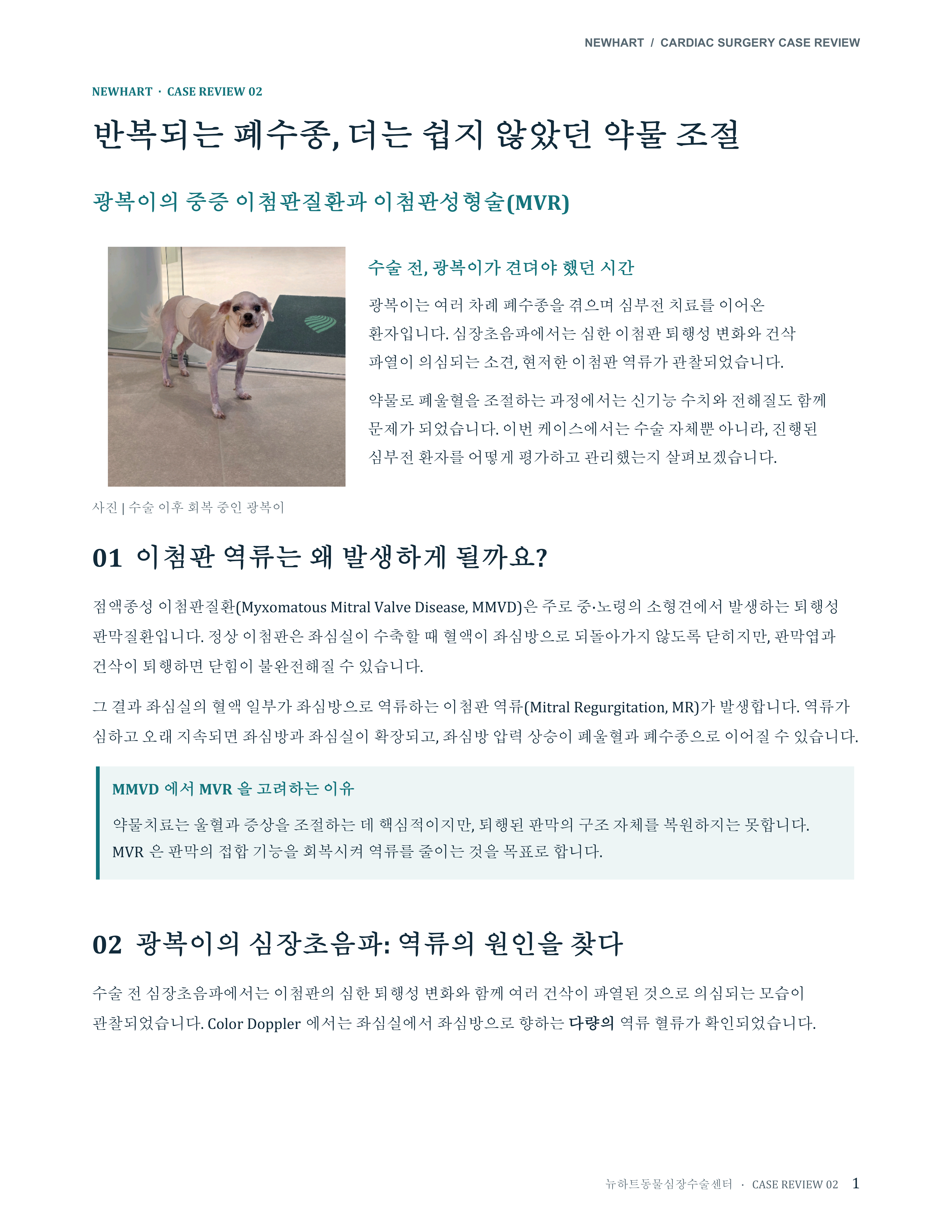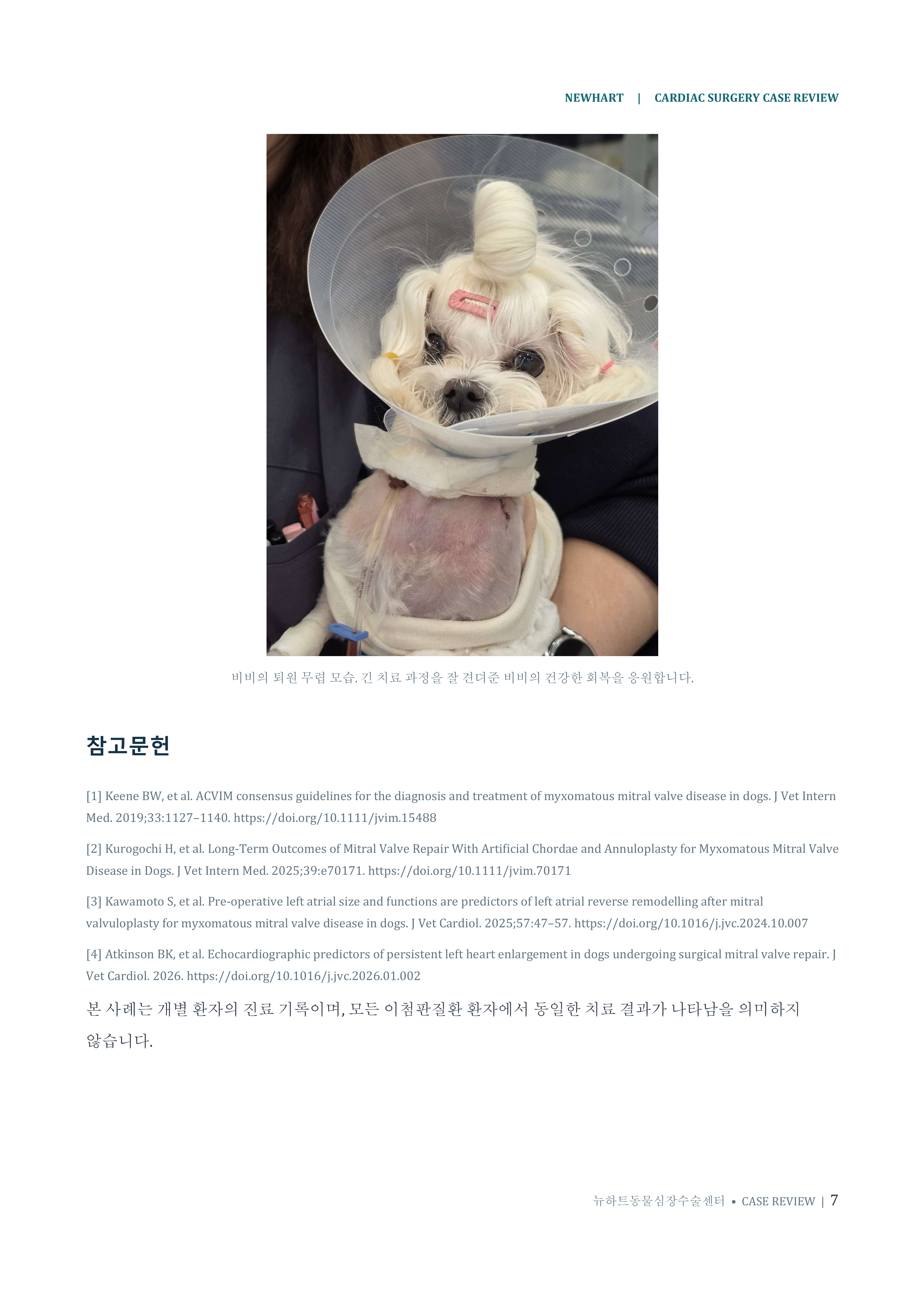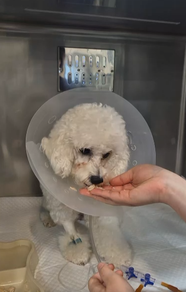

From Hong Kong to South Korea: Odd’s Journey Through Open-Heart Surgery
12 years 5 months old | Severe mitral regurgitation | Extensive chordal rupture | Mitral valve repair (MVR) | Recovery to discharge
뉴하트동물심장수술센터
From Hong Kong to Korea: How 12-year-old Odd found a path toward recovery
Odd, a 12-year-and-5-month-old dog facing severe cardiac disease in Hong Kong, traveled to South Korea for mitral valve repair (MVR). At presentation, persistent respiratory difficulty and profound weakness made it difficult for him even to stand. Echocardiography showed severe mitral regurgitation (MR), together with findings suggestive of pulmonary hypertension.
After assessing Odd’s cardiac and overall condition, the NewHeart surgical team performed open-heart surgery. Extensive rupture of the chordae supporting the mitral valve was identified intraoperatively. Following repair, MR was nearly eliminated, and thoracic radiographs obtained around discharge showed a reduction in the cardiac silhouette.
This case review follows Odd’s journey from Hong Kong to Korea, using actual imaging to illustrate the preoperative findings, intraoperative lesions, postoperative valve function and pulmonary hypertension indices, and recovery through discharge.
01 From Hong Kong to Korea — An Older Patient with Advanced Heart Disease
Odd was 12 years and 5 months old at the time. After receiving a grave prognosis in Hong Kong, his family sought further treatment options and brought him to NewHeart Animal Cardiac Surgery Center.
Upon arrival in Korea, Odd had chronic respiratory difficulty and markedly reduced activity, with difficulty standing independently. His advanced age, severe valvular disease, cardiac enlargement, and findings associated with pulmonary hypertension made both surgery and postoperative recovery particularly challenging.
02 A Mitral Valve Unable to Close — Extensive Chordal Rupture
Preoperative echocardiography demonstrated marked prolapse and a flail appearance of the anterior mitral leaflet, accompanied by extensive regurgitation. The mitral valve normally prevents blood from flowing backward into the left atrium during systole. In Odd, severe damage to the supporting structures prevented adequate leaflet coaptation.
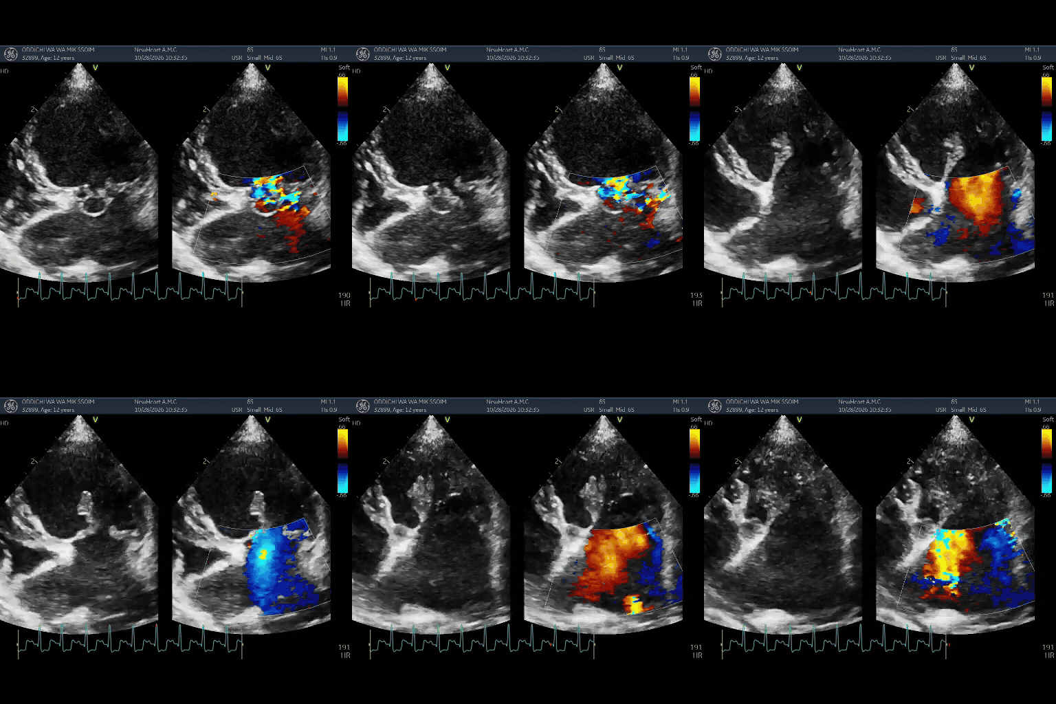
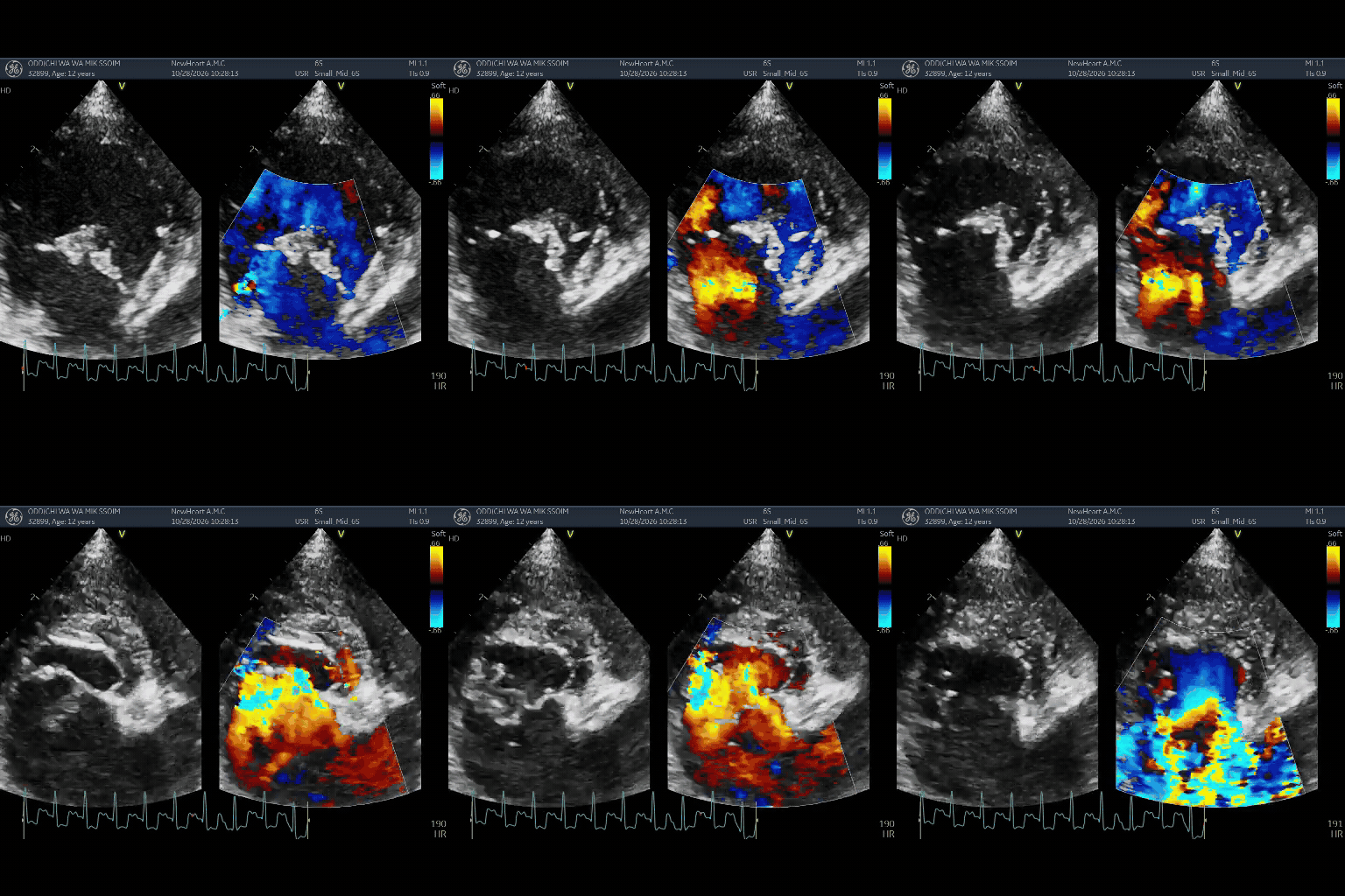
Figures 1–2. Preoperative echocardiography showing severe structural mitral abnormalities and extensive regurgitation.
Intraoperatively, nearly all chordae supporting the anterior leaflet were found to be ruptured, apart from one primary and one secondary chordal structure. This extensive injury was considered the major anatomical cause of the severe MR.
When chordal rupture is widespread, restoring stable valve coaptation becomes technically demanding. The goal is therefore not simply to reduce regurgitant flow, but to reconstruct the compromised support structures and establish an adequate leaflet coaptation surface.
03 Preoperative Findings Suggestive of Pulmonary Hypertension
Before surgery, peak tricuspid regurgitation velocity (TR Vmax) measured 4.55 m/s, with a calculated peak pressure gradient (TR maxPG) of 82.86 mmHg. These findings indicate a high right ventricular–right atrial systolic pressure gradient and strongly support the possibility of pulmonary hypertension.
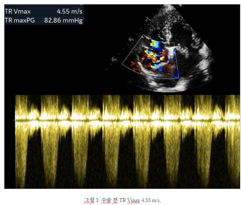
Figure 3. Preoperative TR Doppler: TR Vmax 4.55 m/s and TR maxPG 82.86 mmHg.
Severe MR and increased left atrial pressure can contribute to pulmonary hypertension associated with left-sided heart disease. However, neither the underlying mechanism nor severity of pulmonary hypertension should be determined from a single index; the broader echocardiographic and clinical findings must also be considered.
Thoracic radiographs demonstrated marked enlargement of the cardiac silhouette. The original clinical records also described substantial enlargement of both the left- and right-sided cardiac chambers.
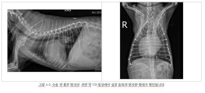
Figures 4–5. Preoperative lateral and ventrodorsal thoracic radiographs demonstrating a markedly enlarged cardiac silhouette.
04 Open-Heart Surgery — Restoring Mitral Valve Coaptation
The NewHeart team performed mitral valve repair using cardiopulmonary bypass. The objective was to reconstruct the support mechanisms damaged by chordal rupture and restore sufficient coaptation of the anterior and posterior mitral leaflets during systole.
Postoperative echocardiography showed markedly improved leaflet coaptation and near-complete resolution of the previously severe MR. Only minimal residual regurgitation was observed in different imaging planes, an important immediate result of the repair.
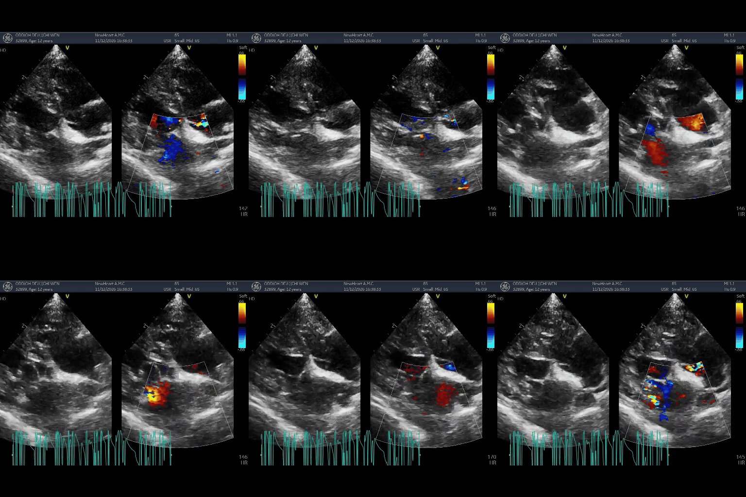
Figures 6. Postoperative echocardiography showing improved mitral coaptation and minimal residual MR in two imaging planes.
These findings indicate successful structural correction at the early postoperative assessment. However, discharge findings alone cannot establish long-term freedom from recurrent MR or predict survival; serial echocardiographic follow-up remains necessary.
05 A Smaller Cardiac Silhouette on Discharge Radiographs
The markedly enlarged cardiac silhouette decreased after surgery. Radiographs obtained around discharge showed a smaller overall cardiac silhouette, with particularly visible changes in the right-sided cardiac contour.
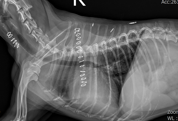
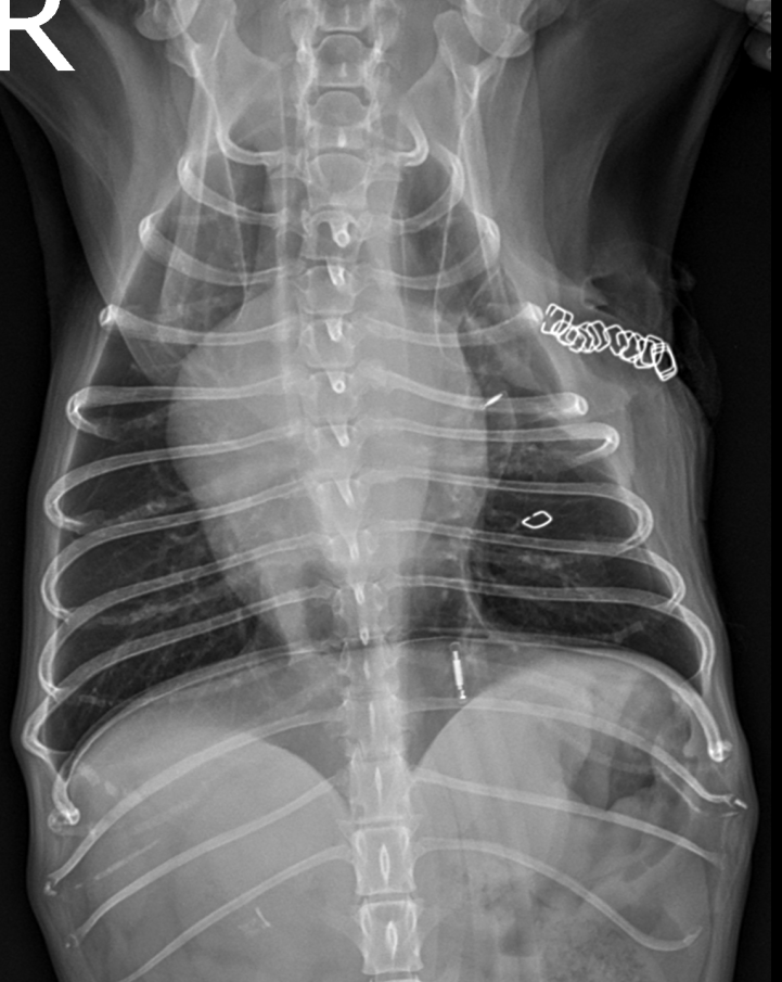
Figures 7–8. Postoperative lateral and ventrodorsal thoracic radiographs showing a smaller cardiac silhouette than before surgery.
This early change is consistent with reduced cardiac volume load after correction of severe MR. Nevertheless, radiographs alone cannot precisely quantify individual chamber dimensions, and sustained reverse remodeling must be assessed through follow-up echocardiography.
06 Pulmonary Hypertension Indicators Remained Elevated
After surgery, TR Vmax decreased to 4.03 m/s and TR maxPG to 64.91 mmHg. Although both measurements were lower than before surgery, the values remained elevated and continued to raise concern for pulmonary hypertension.
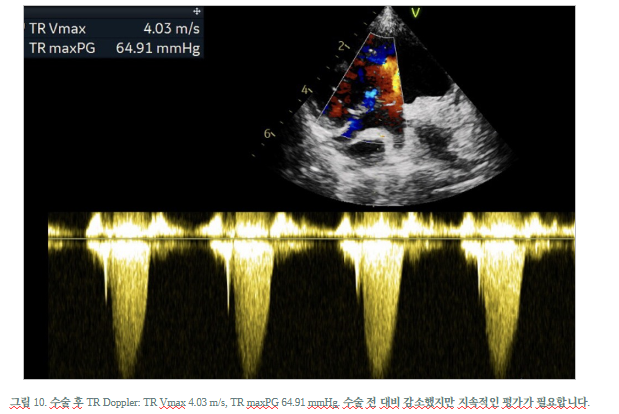
Figure 9. Postoperative TR Doppler: TR Vmax 4.03 m/s and TR maxPG 64.91 mmHg; improved but still requiring follow-up.
Near-complete elimination of MR should not be equated with complete resolution of pulmonary hypertension. Pulmonary circulatory hemodynamics may continue to improve during recovery from sustained left atrial hypertension and recurrent left-sided heart failure, but the extent and timing vary between patients.
Accordingly, follow-up after discharge should address Odd’s respiratory status, pulmonary hypertension indices, right-heart size and function, and response to medication.
07 Odd’s Journey from Hong Kong — Toward Everyday Life Again
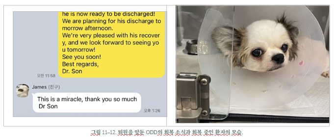
Odd’s surgery brought together his family’s decision to travel from Hong Kong and the NewHeart team’s careful preparation. Although he had been a critically ill older patient unable to stand easily at presentation, he recovered sufficiently for discharge after surgical correction of his severe structural mitral lesion.
Odd’s family expressed heartfelt appreciation for his recovery. We share this case not to suggest that every critically ill patient will have the same outcome, but to demonstrate that careful case selection and coordinated expertise in cardiac surgery, anesthesia, cardiopulmonary bypass, and intensive care can expand treatment possibilities even in advanced structural valve disease.
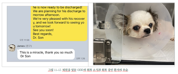
Figures 11–12. Odd during recovery and a message from his family as discharge approached. Any publicly shared correspondence or photographs should be reviewed for consent and personal information.
Discharge marks the beginning of another stage of recovery. Continued follow-up is essential to assess Odd’s long-term valve function and overall cardiac health. Everyone at NewHeart hopes that he will enjoy many more healthy days with his family in Hong Kong.
08 Key Takeaways from This Case
First, extensive chordal rupture was confirmed as an anatomical cause of severe MR, and regurgitation was nearly eliminated after surgical repair. Second, despite marked preoperative cardiomegaly and high TR velocity, both the radiographic cardiac silhouette and TR velocity decreased by discharge. Third, pulmonary hypertension-related indices remained elevated, requiring continued reassessment and treatment as appropriate.
The outcomes described here extend through the peri-discharge period. Suitability for surgery and long-term prognosis vary with disease severity, pulmonary hypertension, renal function, and other comorbidities; individualized clinical evaluation remains essential.
Note: This case review is based on an individual patient’s examinations and surgical records. Imaging comparisons may be influenced by differences in acquisition conditions and timing.
Selected English References | EVIDENCE
[1] Keene BW, et al. ACVIM consensus guidelines for the diagnosis and treatment of myxomatous mitral valve disease in dogs. J Vet Intern Med. 2019;33:1127–1140. doi:10.1111/jvim.15488.
[2] Reinero C, et al. ACVIM consensus statement guidelines for the diagnosis, classification, treatment, and monitoring of pulmonary hypertension in dogs. J Vet Intern Med. 2020;34:549–573. doi:10.1111/jvim.15725.
[3] Kongchatree S, Rattanasrisomporn J. When to consider mitral valve surgery in dogs: a comparative review of open mitral valve repair and transcatheter edge-to-edge repair with a proposed selection framework. Front Vet Sci. 2026;13:1900762. doi:10.3389/fvets.2026.1900762.
[4] Ray CC, et al. Retrospective evaluation of acute postoperative complications occurring in the ICU following canine mitral valve repair surgery under cardiopulmonary bypass (2019–2020): 41 cases. J Vet Emerg Crit Care. 2025;35:736–753. doi:10.1111/vec.70071.
NEWHART ANIMAL CARDIAC SURGERY CENTER
Advanced valve repair, dedicated care throughout recovery
This article is general information and does not replace an individual diagnosis or treatment. If you are worried about your pet, please book a visit.