
One Year After MVR: From Acute Kidney Injury and Severe Pulmonary Edema to Stability
Geoni's 1-Year Follow-up: Surgical Timing and NewHeart's Direct Aortic Cannulation Strategy
뉴하트동물심장수술센터

Geoni was a high-risk patient with severe cardiogenic pulmonary edema complicated by acute kidney injury (AKI). The clinical team first stabilized respiratory and renal status before proceeding with mitral valve repair (MVR) under cardiopulmonary bypass (CPB). At the 1-year follow-up, the left atrium remained small, residual mitral regurgitation was not considered clinically significant, and renal laboratory values were within the reference ranges. This case illustrates how individualized surgical timing and careful perfusion management may support favorable longer-term outcomes in selected patients with prior renal injury.
Geoni at a Glance
01. Initial Assessment: Monitoring After Pulmonary Edema Treatment
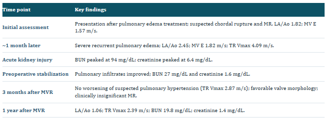
Geoni presented after receiving treatment for pulmonary edema. Echocardiography demonstrated suspected chordal rupture and mitral regurgitation, but the overall findings at that point did not appear severe enough to necessitate immediate surgery.
The left atrium was moderately enlarged (LA/Ao 1.82), and mitral inflow E velocity was 1.57 m/s. Because examination followed a recent episode of pulmonary edema, the team recognized that filling-related indices might change as treatment progressed and planned close reevaluation. The initially recorded TR-derived pressure gradient was approximately 39 mmHg.
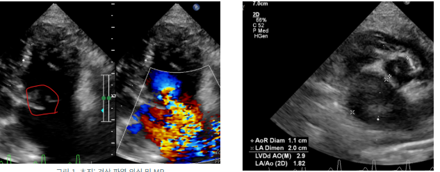
Fig. 1. Initial examination: suspected chordal rupture and MR,
Fig. 2. Initial LA/Ao 1.82
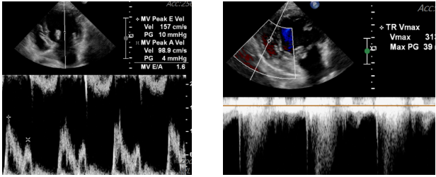
Fig. 3. Initial MV E velocity, 1.57 m/s
Fig. 4. Initial TR gradient, approximately 39 mmHg
02. Sudden Deterioration: Cardiac, Pulmonary, and Renal Stress
Approximately one month later, Geoni returned with acute pulmonary edema. Thoracic radiographs showed marked pulmonary infiltrates, and reevaluation demonstrated substantially worsened mitral regurgitation and rapid left atrial enlargement: LA/Ao increased from 1.82 to 2.45.
Mitral E velocity increased to 1.82 m/s, while TR Vmax reached 4.09 m/s (estimated gradient approximately 67 mmHg), consistent with increased right-sided pressure load at the time of examination. These Doppler measures do not independently establish left atrial or pulmonary arterial pressure, but together with the clinical course they supported a marked hemodynamic deterioration.
Pulmonary edema was difficult to resolve despite treatment. Simultaneously, BUN and creatinine increased sharply, creating a challenging balance between treatment of congestive heart failure and preservation of effective renal perfusion.
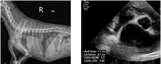
Fig. 5. Acute edema, lateral radiograph
Fig. 6. LA/Ao 2.45 at decompensation
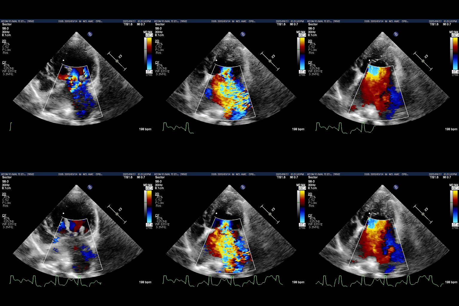
Fig. 7. Worsening MR on color Doppler
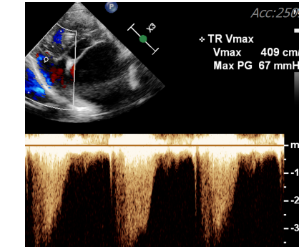
Fig. 8. TR Vmax 4.09 m/s; gradient 67 mmHg
03. Preoperative Stabilization: Timing the Operation Matters
During the acute phase, BUN rose to 94 mg/dL and creatinine peaked at 6.4 mg/dL. The team elected to optimize pulmonary edema treatment and allow as much renal recovery as clinically feasible before MVR.
Serial laboratory measurements subsequently improved. Immediately before surgery, BUN was 27 mg/dL and creatinine was 1.6 mg/dL, while thoracic radiographs documented improvement in pulmonary edema. This did not eliminate operative risk; rather, it reduced modifiable risk factors before instituting CPB.
Geoni demonstrates that severe pulmonary edema and AKI do not necessarily preclude surgery. Instead, the decision requires individual evaluation of disease trajectory, hemodynamic stability, renal recovery, and the hazards of further delay.
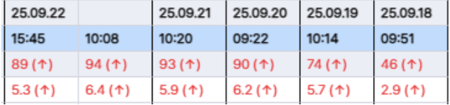
Fig. 9. AKI laboratory trend, September 18–22

Fig. 10. Renal recovery trend through October 2
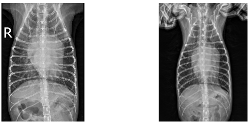
Fig. 11. Persistent pulmonary edema despite treatment
Fig. 12. Improved pulmonary edema before surgery
04. Direct Aortic Cannulation and Systemic Organ Perfusion
During open mitral valve repair, the CPB circuit must provide systemic blood flow while the heart is temporarily arrested. In a patient with a recent history of AKI, the adequacy of pump flow and arterial pressure, oxygen delivery, anemia and hemolysis, temperature management, and postoperative hemodynamics are especially important.
NewHeart utilizes direct aortic cannulation to establish arterial inflow into the descending aorta, rather than routing inflow through a carotid artery. In an abstract presented at the 2026 ACVS Surgery Summit, the NewHeart team described this approach as permitting a larger cannula and standard-flow perfusion under moderate hypothermia while preserving both carotid arteries.
Direct cannulation of the aorta, by itself, has not been shown to protect the kidneys. Nevertheless, a reliable arterial inflow strategy that supports adequate CPB perfusion is physiologically relevant to efforts to limit renal hypoperfusion, low oxygen delivery, and hemodynamic instability. Geoni's favorable renal course is consistent with the broader goal of carefully managed systemic perfusion, but it cannot establish the independent protective effect of the cannulation site.
05. Three Months After Surgery: Improved Valve Function
At the 3-month postoperative examination, thoracic radiographs showed a favorable cardiopulmonary appearance. Echocardiography demonstrated a stable repaired mitral valve configuration, with residual MR described as clinically insignificant.
TR Vmax decreased from 4.09 m/s at the acute deterioration visit to 2.87 m/s. This supports a lower TR-derived pressure gradient under the conditions of the follow-up examination; there was no imaging evidence of worsening pulmonary hypertension at that visit.
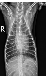
Fig. 13. Three-month thoracic radiograph
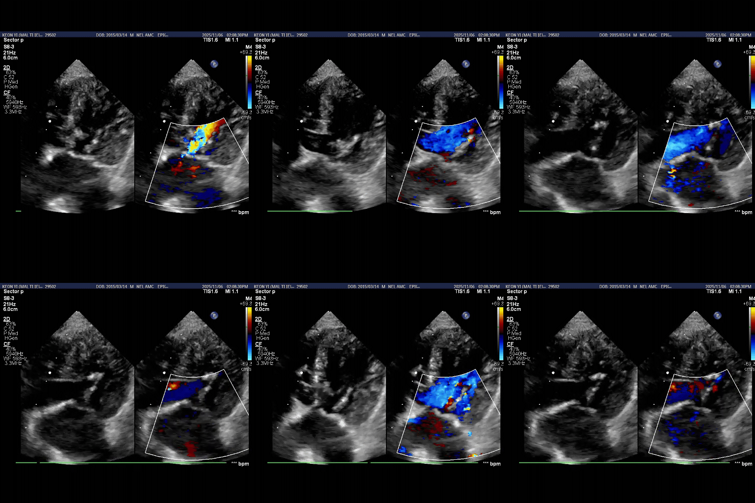
Fig. 14. Three-month valve structure and MR
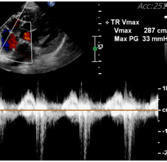
Fig. 15. Three-month TR Vmax 2.87 m/s
06. One Year After Surgery: Sustained Cardiac and Renal Stability
At one year, thoracic radiographs revealed no new significant cardiopulmonary abnormalities. Echocardiography showed a non-enlarged left atrium with LA/Ao 1.06. Maintaining this degree of reverse remodeling after the marked preoperative enlargement is a notable long-term observation.
TR Vmax was 2.39 m/s (estimated gradient 22.8 mmHg). Renal laboratory values were within the reported reference intervals: BUN 19.8 mg/dL and creatinine 1.4 mg/dL. The available one-year results provided no evidence of progressive azotemia.
These findings show that a selected dog with a history of severe AKI can maintain favorable cardiac and renal status after careful preoperative stabilization, CPB management, and successful valve repair. The case documents recovery from AKI, however, rather than proving safety for every patient with advanced chronic kidney disease (CKD).
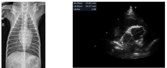
Fig. 16. One-year thoracic radiograph
Fig. 17. One-year LA/Ao 1.06
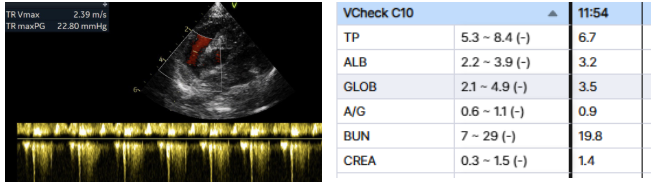
Fig. 18. One-year TR Vmax 2.39 m/s
Fig. 19. One-year BUN 19.8; creatinine 1.4 mg/dL
07. The Clinical Message: Treating the Heart Without Losing Sight of the Kidneys
Geoni's outcome cannot be attributed to valve surgery alone. The team first chose a surgical window after the acute respiratory and renal deterioration had improved. A carefully planned CPB arterial inflow strategy and attentive perioperative management were then central elements of the treatment approach.
NewHeart's direct aortic cannulation technique is one component of that strategy. Although the independent renal benefit of the cannulation route cannot be determined from a single case, the technique highlights the importance of planning for reliable systemic organ perfusion in patients whose renal reserve has already been challenged.
One year after MVR, Geoni maintained a small left atrium, clinically insignificant residual MR, and renal values within the laboratory reference ranges. A history of renal injury does not automatically mean that open-heart surgery is impossible. With individualized risk assessment, appropriate timing, and attentive perfusion management, treatment options may remain available for carefully selected patients.
References and Technical Background
[1] Eom T. Direct Aortic Cannulation for Canine MVR. Scientific abstract, 10 October 2026. https://surgerysummit2026.eventscribe.net/fsPopup.asp?PresentationID=1863396&mode=presInfo
[2] Retrospective Evaluation of Acute Postoperative Complications Following Canine MVR Under CPB: 41 Cases. J Vet Emerg Crit Care. 2025;35:736–753. doi:10.1111/vec.70071. https://pmc.ncbi.nlm.nih.gov/articles/PMC12779198/
[3] Cardiopulmonary bypass associated acute kidney injury: better understanding and better prevention. Ren Fail. 2024. doi:10.1080/0886022X.2024.2331062. https://pmc.ncbi.nlm.nih.gov/articles/PMC10962309/
[4] Kamla C-E. Kidney Injury Following Cardiac Surgery: A Review of Our Current Understanding. Drugs. 2025;85:337–348. doi:10.1007/s40256-024-00715-8. https://pmc.ncbi.nlm.nih.gov/articles/PMC12014718/
Values are drawn from the provided clinical images and laboratory summaries. Doppler-derived pressure gradients are estimates. Continuing cardiac and renal follow-up remains important when interpreting the long-term prognosis.
NewHeart Animal Cardiac Surgery Center · NEWHART CASE REVIEW
This article is general information and does not replace an individual diagnosis or treatment. If you are worried about your pet, please book a visit.